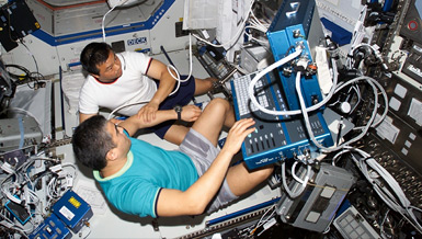
Although the effects of space flight on the immune system are linked to the weightless environment, mechanisms behind these changes are not understood. Dr. Yufang Shi is examining the molecular mechanisms involved in the alteration of the immune system during space flight. He also will test the hypothesis that the immune system contributes to bone loss under weightless conditions.
Overview
Effects of Antiorthostatic Suspension on the Immune System
Principal Investigator:
Yufang Shi, D.V.M., Ph.D.
Organization:
University of Medicine and Dentistry of New Jersey
Robert Wood Johnson Medical School
Technical Summary
Space flight causes lymphopenia and suppression of the immune system in humans, monkeys, and rodents - lymphocyte numbers, mitogen response and production of cytokines and antibodies are all significantly reduced. Several factors including microgravity, lack of load-bearing, stress, acceleration forces and irradiation likely contribute to these changes, but the exact mechanisms are unknown. Simulation of these factors on the ground using animal models such as antiorthostatic suspension (now termed hindlimb unloading (HU), results in similar immune system changes. We hypothesized that immune system changes during space flight are modulated through Fas in an endogenous opioid-dependent manner.
Our specific aims are:
- Investigate the modulation of the Th1 and Th2 responses in mice subjected to HU;
- Explore the mechanisms of HU-induced thymus involution, and;
- Examine the role of RANKL in the communication between the immune and skeletal systems during HU.
Key Findings
We have completed the characterization of lymphocyte phenotypes from mice subjected to HU. We found that not all mouse strains are equally susceptible to HU, with Balb/c being most sensitive and C3H the least, indicating genetic variability in susceptibility to HU. Nevertheless, splenocyte and thymocyte numbers decreased significantly in all mouse strains after HU. This effect occurs through apoptosis, evidenced by genomic DNA fragmentation and phosphatidylserine exposure (TUNEL staining). Interestingly, splenocyte loss required endogenous opioid-mediated Fas expression; the effect in thymus was corticosteroid-dependent. Thus, the dramatic effect of HU on lymphocyte numbers is exerted through different mechanisms in spleen and thymus.
We have begun to develop countermeasures against HU-induced lymphocyte loss. Since we found that free-radicals are important in the apoptosis process in vitro, we tested the effect of free-radical scavengers. Vitamin C, a common food supplement with potent scavenger capacity, was tested for its ability to prevent HU-induced lymphocyte apoptosis. Balb/c mice were given vitamin C in drinking water (two mg/ml) for ten days then subjected to HU for two days. Lymphocytes in spleen and thymus were enumerated, and subsets examined by flow cytometry, splenocyte and thymocyte numbers were reduced by about 50 percent following HU vs. untreated controls.
CD4+CD8+ double-positive thymocytes were particularly vulnerable. Vitamin C protected the thymus but not the spleen, and double-positive thymocytes were the most significantly protected. The lack of protection in spleen may reflect a different mechanism of HU-induced lymphocyte depletion in this organ.
Another promising countermeasure is seaweed polysaccharide extract. Countermeasures against radiation- or stress-induced apoptosis have been vigorously pursued, yet no safe, effective reagent has been found for human use. We tested a polysaccharide extract from brown seaweed (part of the Eastern diet for centuries) using a commercial product from NaturoDoc. When fed at 100 mg/mouse/day for five days, this extract significantly inhibited radiation-induced splenocyte loss. Such an extract may be a safe and effective countermeasure against radiation-induced immune system damage for astronauts.
Bone loss is a major concern for astronauts. Blockade of RANKL with OPG is known to inhibit HU-induced bone loss. We hypothesized that corticosteroid production mediates this process. We found that corticosteroid-induced RANKL expression in T cells depends on the mobilization of intracellular calcium. Surprisingly, free-radicals are also required, pointing to potential countermeasures.
Efforts to elucidate the role of CD4+CD25+ T cells (Treg) in HU-induced lymphocyte loss continue. We have found that Treg resist depletion by HU, while artificial depletion of Treg restores antigenic T cell responses in vitro. Conditions for the effective depletion of Treg in vivo have been established, and genetically modified mice were chosen to examine how Treg are involved in lymphocyte depletion.
Research Plans
We will continue to develop countermeasures for HU- and radiation-induced lymphocyte reduction. We will further examine the mechanisms by which vitamin C is protective. We found that vitamin C inhibits the effect of dexamethasone in vitro, so we will use this system to look for an effect on RAGE and BIM, two molecules implicated in thymocyte apoptosis. The relationship between vitamin C and free-radicals will also be examined.
Our studies with polysaccharide extract from brown seaweed are promising. At present, we have no information about the mechanisms of its protective effect. We will determine if the extract protects cells from radiation in vitro to establish a direct effect on cells. If this is not the case, we will examine the effect of serum from mice fed with the extract. In addition, mice fed with or without the extract and irradiated will be compared for apoptosis, DNA damage, free-radical production and survival. The extracts effect on HU-induced lymphocyte reduction will also be tested. Meanwhile, we will collaborate with Dr. Ann Kennedy to test the effect of a diet developed in her lab on HU-induced lymphocyte changes. We will complete studies on the role of calcium and free-radicals in corticosteroid-induced RANKL expression. A manuscript describing these results will be submitted soon.
We have found clear changes in the production of IL-4 and IFN-gamma in mice subjected to HU. These experiments have relied on ELISA, a slow and tedious way to assay multiple cytokines. We recently installed the Bio-Plex (Luminex) multiplexed microbead assay system, capable of detecting up to 28 cytokines at once. We will use it to systematically assay multiple cytokines in serum from mice immunized with ovalbumin during HU-treatment. This will provide us a broader understanding of the effect of HU on the immune system.
Earth Applications
- Understanding the mechanisms by which stress affects immune homeostasis may lead to the identification of dietary and lifestyle changes that reduce the deleterious effects of stress on the immune system. Adopting such changes may lead to improved public health. In addition, we may identify food supplements such as vitamin C and seaweed extracts that minimize the impact of stress, thus benefiting the general population, especially those who suffer from high stress levels occupationally or due to life circumstances.
- Learning the effects of manipulating a specific subset of T lymphocytes called CD4+CD25+ regulatory T cells may lead to potential interventions to prevent the deleterious health effects of autoimmunity, cancer and other diseases involving over- or under-activity of the immune system.
- Identification of a protein(s) whose serum concentration is related to the stress levels experienced by an individual may provide a marker to quickly and accurately assess stress levels in medical or occupational contexts.
- Elucidation of the role of cellular apoptosis in mediating the deleterious effects of stress on the immune system may aid in the development of clinical protocols for maintaining immune tolerance such is in transplantation or autoimmune disease.


