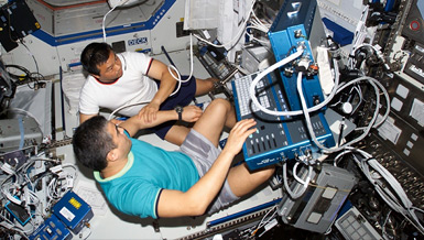
When a person fractures a bone on Earth, the process to heal the bone is effective in most instances. Previous research indicates that fracture healing could be impaired on the lunar surface, where gravity is one-sixth of Earth’s. Dr. Ronald J. Midura leads the continuation of his previous project. He is seeking to determine how much bone healing is impaired and what causes the impairment. During this animal study, he will also develop countermeasures to prevent fracture healing problems and will determine if Earth-based clinical procedures are effective in reducing the effects of reduced gravity on fracture healing.
Overview
Extent, Causes and Countermeasures of Impaired Fracture Healing in Hypogravity
Principal Investigator:
Ronald J. Midura, Ph.D.
Organization:
The Cleveland Clinic
Technical Summary
The healing of fibular fractures in rats during actual spaceflight or under simulated hypogravity conditions is deemed to be impaired. This project seeks to continue research which found that fibular osteotomy healing in hindlimb unloaded (HLU) rats was delayed, leading to a significant number of non-unions, and was associated with a substantially reduced number of marrow-derived osteoprogenitor cells providing a partial explanation for impaired healing. Also, bone anabolic drugs decreased the incidence of fibular non-unions and improved the number of osteoprogenitor cells. Altogether, this suggests that fracture healing in space is not Earth normal and provides the rationale to further investigate whether impairment of fibular fracture healing would extend to more clinically relevant closed femoral fractures. Our global hypothesis is that long-duration hypogravity impairs fracture healing.
Objectives
- Determine the scope and extent of femoral fracture healing impairment.
- Determine the underlying biological causes of the impairment.
- Develop countermeasures to prevent fracture healing impairment.
- Determine whether current Earth-based clinical procedures will reverse severely delayed fracture healing situations resulting from hypogravity.
- Micro-CT bone imaging to evaluate hard callus structure;
- Hard callus strength via torsion testing;
- Callus tissue composition using histomorphometry;
- Colony forming unit assessments of marrow-derived osteoprogenitor cell numbers; and
- Measurements of osteoinductive, chondrogenic and angiogenic factor expression during early healing periods.
In its second year, the project confirmed the initial findings that closed femoral fractures in HLU rats exhibit substantially smaller hard callus volumes (40-60 percent smaller than WB ones) even after 10-weeks of healing. Yet, torsion testing assessments of HLU vs. WB hard calluses indicated sound mechanical properties for both HLU and WB calluses, though the HLU calluses were more brittle. Histological assessments at 10-weeks indicate that the content within the HLU calluses is about 40% mineralizing tissue and about 20% soft/fibrous tissue. Assessments of gene expression and tissue alterations for early fracture healing time points (one and two weeks post-fracture) correlating to the chrondrogenic phase (soft tissue callus formation) and the beginnings of the endochondral ossification phases (hard tissue callus formation) of fracture healing are complete. This analysis highlights a delay in endochondral ossification due to lagging chondrocyte hypertrophy, in effect delaying subsequent steps of the healing process such as angiogenic vessel infiltration and mineral deposition. Safranin O histological staining results of proteoglycan deposition within the fracture callus from WB and HLU rats suggested similar amounts of hyaline cartilage tissue in each test group. These histological findings are in agreement with those of gene expression findings whereby aggrecan mRNA levels were similar between groups at one and 2 weeks post-fracture. This prolonged chondrocyte maturation step exemplified by delayed hypertrophy, reduced osteo-inductive factor expression, and reduced pro-angiogenic factor expression likely leads to a postponement of the requisite vascularization of HLU callus tissue and its subsequent mineralization. While after a full 10 weeks of healing, it is apparent that HLU fractures heal, the data also indicate that the healing process itself may be altered as compared to fractures from WB rats. In fact, fractures from HLU rats are mechanically sound compared to fractures from WB rats. Yet the explanation for this adaptive healing response in HLU callus is not identified currently.
In its third year, the project has utilized pharmaceuticals and biophysical stimulation in attempts to augment fracture healing in rat femora. Both WB and HLU rats were given intermittent parathyroid hormone (PTH) injections to stimulate fracture healing and fractures were monitored longitudinally by micro-CT at multiple post-fracture time points. Analysis of this data is ongoing. Low intensity pulsed ultrasound (LIPUS) was also utilized to augment fracture healing in HLU rats. Preliminary findings indicate that fracture callus bridging occurs more rapidly in response to LIPUS than to sham treated femoral fractures. In addition to these findings, further analysis of fracture callus after 10 weeks of non-pharmaceutical or -biophysical stimulated healing by histomorphometrics confirms micro-CT data indicating that total callus volumes are decreased in HLU fractures and that HLU callus tissue contains reduced amounts of cartilagenous and fibrous tissues. Use of anti-sclerostin as a pharmaceutical method to augment healing has not been completed as yet due to delays in obtaining the antibody. We anticipate continuing this study as part of year 4 work.
Earth Applications
The impact of our findings for Earth-based medical practice would suggest that an extended period of unloading and a cephalic fluid shift out of normally weight-bearing lower extremity bones may manifest a delayed or an impaired bone healing response. This information may have relevance toward a better understanding of the underlying causes of impaired bone healing in patients experiencing paralysis, chronic immobility or extended bed rest. Previous data suggested that treatments with bone anabolic therapies seem to partially counteract the impairment of bone healing under simulated spaceflight conditions. This project will explore additional potential countermeasures in the third year and may also offer potential treatments for augmenting bone healing in Earth-bound, non-weight-bearing patients.


