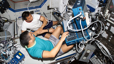
Astronauts exposed to microgravity conditions have been noted to develop a spectrum of neurologic and ophthalmic disorders, including symptoms of idiopathic intracranial hypertension and papilledema. Translaminar pressure gradients between the intraocular and intracranial spaces, with the latter demonstrating elevated pressure, are postulated to be responsible for some of the ocular symptoms associated with prolonged space travel. Methods for early detection of elevated intracranial pressure while the astronaut is still traveling would enable decision-making regarding treatment of the astronaut in space versus return to earth before disease progression.
Current algorithms for diagnosis of elevated intracranial pressure may include a specialist’s physical examination, brain-imaging studies such as MRI (magnetic resonance imaging), and direct measurement of pressures in spaces contiguous with the subarachnoid space using invasive means such as a needle tap or placement of a pressure monitor. None of these algorithms is easily performed in space. Less invasive experimental methods under development include several methodologies that assess quantitative parameters that are not physiologic. For example Doppler based techniques assess cerebral blood flow and ultrasound based techniques assess brain density. These methodologies have the limitation of not assessing the physical consequences of elevated ICP. We have developed a novel method utilizing eye movement tracking technology to quantitate the function of the four cranial nerves responsible for vision and ocular motility. The method consists of having a subject watch a 220 second video (e.g. cartoons or a favorite television show) as it moves in a set rectangular trajectory, while a camera records their eye movements.
Double vision and other ocular disturbances associated with elevated intracranial pressure were first described by Hippocrates in approximately 400 B.C.. Papilledema, and its association with elevated intracranial pressure (ICP) was described by Albrecht von Graefe in 1860. In the post-radiographic era, acute and chronic pathology of the optic nerve and disc, and of ocular motility are well characterized in hydrocephalic children and may precede computed tomography (CT) findings. Several potential mechanisms may contribute to cranial nerve dysfunction due to elevated intracranial pressure. The IIIrd nerve (oculomotor) may be directly compressed by the medial aspect of the temporal lobe with frontal or temporal mass lesions, or diffuse supratentorial mass effect. The VIth nerve (abducens) is anatomically vulnerable to infratentorial mass effect at the prepontine cistern and to hydrocephalus from stretch as it traverses the tentorial edge. Elevated ICP slows axoplasmic transport along cranial nerves. The optic nerve (II) is most frequently analyzed because it can be visualized directly with ophthalmoscopy, and indirectly with ultrasound. Edema of the optic nerve appears earlier than ocular fundus changes, and resolves after treatment of elevated ICP. Fluctuating elevated neural pressure leads to impaired axonal transport along the optic nerve after as little as 30 minutes in a rabbit model. Axoplasmic flow stasis and intraneuronal ischemia may occur in the optic nerve exposed to chronically elevated ICP. Papilledema, or swelling of the optic disc apparent on ophthalmoscopic examination, may be a relatively late indicator of elevated ICP. In one study papilledema was present in as few as 14% of patients with a shunt malfunction consistent with the relatively short intracranial course of II compared to cranial nerves III and IV. Compartmentalization of subarachnoid spaces is hypothesized to explain why papilledema may be present in a patient without elevated ICP, and not occur in patients with elevated ICP.
In order to assess ocular motility including the function of III, IV, VI and associated nuclei, we developed a novel technique for automated eye movement tracking. The position of the pupil is predicted based on time elapsed since the start of the video rather than spatial calibration, enabling detection of impaired ability to move the pupils relative to each other over time. Because this eye tracking technique assesses the function of cranial nerves that are impacted by acute changes in intracranial pressure, it is extremely sensitive to elevations in ICP. It offers the additional advantage of being able to be performed remotely, automatically, and theoretically, during tasks such as reading, without the awareness of the subject being tracked.
To determine the utility of this eye tracking algorithm for detection of elevated ICP during space travel, one must first assess its efficacy on earth. To that purpose we propose two specific aims.
Specific Aim 1: To determine eye tracking metrics associated with measured intracranial pressure (ICP) values in patients with brain injury and other lesions (e.g., hydrocephalus) causing elevated pressure admitted to the ICU for invasive neuromonitoring.
Method: we will recruit 30 subjects who are awake, capable of watching television and have had a ventriculostomy catheter placed for any reason (such as trauma, hydrocephalus, subarachnoid hemorrhage, tumor resection or intracranial hemorrhage). We will eye track these subjects prior to catheter placement (if possible), but otherwise at daily intervals while the catheter is draining or clamped for attempted weaning. We will compare intracranial pressure values at the time of tracking to 51 eye tracking metrics. These metrics reflect the function of cranial nerves II, III and VI, all of which are susceptible to elevated ICP.
Hypothesis: Metrics associated with cranial nerve VI palsy will be the first to be associated with elevated intracranial pressure. Metrics associated with cranial nerve II palsy will only be associated if there is prolonged elevated pressure. Metrics associated with cranial nerve III will only be associated if there is concordant supratentorial mass effect.
Subaim: To assess whether any recruited subjects with papilledema have alterations in eye tracking metrics associated with optic neuropathy.
Method: We will consult ophthalmology for funduscopic examination of any recruited subjects with suspected papilledema. Eye tracking metrics of subjects with papilledema will be compared to themselves when they are recovered and also to subjects without papilledema.
Metrics specifically associated with papilledema on eye tracking are those that reflect optic neuropathy (y-variability and skew).
Specific Aim 2: To demonstrate that subjects with mass effect (swelling) causing narrowing of the space adjacent to the third cranial nerve (perimesencephalic cistern or PMC) as assessed by volumetric analysis of CT (computed tomograph) scan imaging have decreased vertical eye movement on tracking.
In order to benefit astronauts, whose pathology more closely resembles idiopathic intracranial hypertension than diseases requiring invasive intracranial pressure monitoring, an additional goal of this project is to make it possible to correlate eye movement metrics to MRI metrics without having to have an intracranial pressure monitor present in the patient. When exposed to elevated intracranial pressure, the volume of the space around the brainstem (the cerebrospinal fluid containing perimesencephalic cistern) is decreased19. The third nerve runs in/near that space and thus should be under pressure and result in changes in eye tracking. In order to make this correlation we must show that eye tracking metrics are affected when perimesencephalic cistern volume is decreased as a result of elevated ICP.
Method: The same 30 subjects who are recruited for Aim 1 who are awake, capable of watching television and have had a ventriculostomy catheter placed for any reason (such as trauma, hydrocephalus, subarachnoid hemorrhage, tumor resection or intracranial hemorrhage) will have undergone serial CT scans as part of their standard of care. We will volumetrically analyze these scans to quantitate the volume of the perimesencephalic cistern. We will compare cistern volumes at the time of tracking to 51 eye tracking metrics. These metrics reflect the function of cranial nerves II, III and VI, all of which are susceptible to supratentorial mass effect.
Hypothesis: Metrics associated with cranial nerve III palsy will be most strongly associated with supratentorial mass effect as quantitated by decreased PMC volume.


