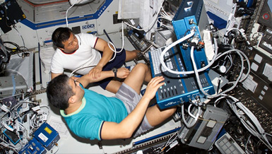
Diagnosing and monitoring medical problems during long-duration flights will be critical to mission success, but these tasks must be carried out by astronauts with limited medical training and equipment. Near infrared spectroscopy (NIR) can noninvasively measure oxygen levels, pH and red blood cell count by using light that passes through the skin and is reflected back to a sensor. However, NIR is not accurate for people with dark skin and significant fat content. Dr. Babs R. Soller is developing a new NIR device to measure blood and tissue chemistry for all humans. Since NIR does not impact blood levels, it allows multiple measurements for continuous monitoring and feedback on medications and other therapeutic measures. This technology can be used on spacecrafts, emergency vehicles and in hospitals.
Overview
Noninvasive Measurement of Blood and Tissue Chemistry (2001-2004)
Principal Investigator:
Babs R. Soller, Ph.D.
Organization:
University of Massachusetts Medical School
Technical Summary
Specific Aims
- Develop calibration procedures for modified NIRS sensor to measure deep muscle metabolic parameters (tissue pH and oxygen);
- Validate sensor in exercise protocol and deliver systems to NASA JSC;
- Determine values for tissue pH and oxygen that can be used by a smart medical system to indicate shock and assist in guiding treatment; and
- Evaluate hardware for flight requirements and develop plan to produce flight-ready instrumentation.
The phantoms and Monte Carlo model helped us identify key sensor design issues to assure optical depth penetration through fat on the forearm and thigh. One system was optimized for thinner fat layers and was made available to NASA JSC for handgrip studies and measurements on the calf during treadmill exercise. We redesigned this sensor and the accompanying monitor to achieve good muscle spectra through thick layers of fat overlying the thigh muscle. One of these systems was delivered to NASA JSC for thigh measurements during treadmill studies.
A new method for calculating muscle oxygen saturation (SmO2) was developed, and its precision was determined as part of a handgrip study at NASA JSC. We also worked with NASA JSC to demonstrate that we could use the pH equation to noninvasively determine hydrogen ion threshold as a surrogate for lactate threshold during cycle ergometry.
Another goal of our project was to develop noninvasive methods to measure absolute values for key parameters so they can be used as part of a smart medical system to help diagnose and guide treatment for critical injuries. To do this, we needed to establish normal values for new parameters and be able to identify values of these new parameters that indicate when someone is sick or getting better. Working with the U.S. Army, we used lower-body negative pressure as a model to simulate the early stages of hemorrhagic shock or internal bleeding. We demonstrated specific values for SmO2 and PmO2 as early indicators of internal bleeding and showed that they provided significantly earlier warning than currently used clinical parameters.
We also conducted a study on sepsis patients undergoing resuscitation with the standard clinical protocol Early Goal Directed Therapy. It showed that SmO2 could be used to indicate when a patient was under-resuscitated while noninvasively determined muscle pH indicated when patients were over-resuscitated with chloride-containing solutions which cause acidosis. Specific values for these parameters were established to provide noninvasively determined goals to direct treatment of septic patients.
We have developed and demonstrated accurate methods for determining muscle oxygen and pH for sick and healthy individuals independent of their skin color and fat thickness. We have demonstrated that these methods can be used on exercising individuals and have applicability for very sick patients being treated in the emergency room. These advances position us to apply a combination of these parameters for determining oxygen consumption during extravehicular activity and assessing muscle and aerobic deconditioning during space exploration.
The NIRS noninvasive metabolic monitor is expected to have many applications for NASA. The system will have additional use on Earth for military and civilian personnel treating critically ill and injured patients. It can also be used in the hospital, ambulances and helicopters. As part of a smart medical system, advanced medical assessment and monitoring may become available to physicians in remote and rural areas, who may not have access to specialist expertise.


