


Astronauts on long lunar missions will face the adverse affects of microgravity, reduced gravity and radiation exposure. It is known that microgravity causes bone loss since bones of the lower body do not bear weight in space like they do on Earth. The impact of radiation on bone quality and fracture healing in reduced gravity is unknown. Dr. Ted A Bateman is investigating the effect of different types of space radiation on bone to learn whether radiation increases the rate of loss. His team is also testing the protective effects of pharmacological countermeasures, such as bisphosphonates, antioxidants and certain proteins.
Overview
Space Radiation and Bone Loss: Lunar Outpost Mission-Critical Scenarios and Countermeasures
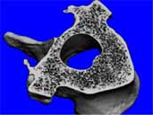
A vertebra from a sheep. Click here for larger image.
Principal Investigator:
Ted A. Bateman, Ph.D.
Organization:
University of North Carolina at Chapel Hill
Technical Summary
Crews on exploratory missions will face complex radiation from cosmic and solar sources with components ranging from protons to iron. We have identified trabecular bone loss in mice after exposure to multiple radiation types with doses ranging from 0.3 Gy to 2 Gy, suggesting space radiation may increase bone loss from reduced gravity during exploratory missions. The bone loss is rapid and initiated by an early activation of osteoclasts.
The impact of radiation on bone quality and fracture healing in reduced gravity is unknown, and must be studied to understand effects of space radiation on bone health. The long-term objective of the proposed research is the development of countermeasures to prevent bone loss during missions and thus reduce fracture risk.
Specific Aims
To define the risks associated with space radiation-induced bone loss, the following aims were studied to examine the effects of modeled space radiation using scenarios applicable for Lunar Outpost missions:
1) Examine the combined effects of a modeled solar particle event and unloading on bone, and subsequent recovery during reloading. Hypothesis: Proton radiation with unloading will induce a more severe bone loss than unloading alone.
2) Examine the cellular and molecular mechanisms for initiating bone loss following exposure to several types of modeled space radiation, including acute proton exposure, low-dose-rate proton exposure, and mixed radiation types (proton and HZE). Understanding underlying molecular causes is critical to developing countermeasures for radiation-induced bone loss. Hypothesis: The initiating mechanism of bone loss is initiated by osteoclast activation caused by a radiation-induced inflammatory response.
3) Test the efficacy of three countermeasures for bone loss caused by proton exposure: the
bisphosphonate risedronate, the RANKL blocking protein osteoprotegerin, and an antioxidant agent, alpha-lipoic acid.
Summary of Aim 1 Findings
• Mice that were exposed to a 1 Gy dose of protons and 28 days of hindlimb suspension experienced damage that was additive. Irradiation caused a 15% decline in trabecular bone mass in normally loaded mice and a 17% decline in skeletally unloaded mice.
• A long-term suppression of bone formation inhibits recovery of bone mass.
Future Direction of Aim 1: The effects of mixed radiation, modeling solar particle events and cosmic radiation, needs to be modeled with fractionated exposure during skeletal disuse. It is important to understand if skeletal disuse modifies the threshold dose at which bone loss from radiation is observable. The degree to which animal models predict the human response to space radiation (astronauts) needs to be better understood by studying cancer patients receiving radiation therapy, particularly at the periphery of the radiation field where doses are lower.
Summary of Aim 2 Findings
• The acute response to 50 cGy dose of protons or heavy ions is the activation of osteoclastic bone resorption.
• Serum markers for bone resorption increase 24 hours after exposure and osteoclast numbers and eroded surface are greater at thee days.
• Osteoclast activation is temporary, reducing to baseline levels approximately 10-days after irradiation.
• The researchers identified that this bone loss is preceded by death of bone marrow cells, followed by greater expression then levels of inflammatory cytokines IL-1, IL-6 and TNFalpha.
• This osteoclast-mediated acute response is not mitigated by low-dose-rate exposure to protons.
• Acute, osteoclast-mediated bone loss from 50 cGy proton exposure is approximately 50% that of a 2 Gy exposure.
• The acute bone loss caused by heavy ions (oxygen, silicon, neon, iron) is approximately 30% - 50% greater than that caused by protons.
Future Direction of Aim 2: A more detailed description of acute relative biological effectiveness (RBE), both at the functional and cellular level, needs to be characterized. The association of acute marrow death and osteoclast activation needs to be studied at the causation level with cell culture models.
Summary of Aim 3 Findings
• The osteoclast inhibiting bisphosphonate risedronate fully prevented bone loss from a 2 Gy whole body dose of X-rays.
• Because of intellectual property concerns with a patent application filing, the RANKL-inhibiting protein.osteoprotegerin was not able to be tested.
• As a substitute for OPG, two additional bisphosphonates, pamidronate and zoledronate, were tested..Zoledronate was more effective than pamidronate.
• Neither of the antioxidant compounds alpha-lipoic acid nor n-acetal cystine (NAC) reduced bone loss.from radiation exposure.
• The IL-1 and TNFalpha blocking compounds, IL-1ra (Kineret) and TNFbp (Enbrel), were tested as.potential countermeasures. Neither protein, independently or in combination, mitigated bone loss from.radiation exposure.
Future Direction of Aim 3: As an antiresorptive agent that suppresses osteoclast activity through a different mechanism than bisphosphonates, osteoprotegerin should be studied as a countermeasure (intellectual property concerns are currently being addressed). The most effective dosing regimen for risedronate and zoledronate needs to be be further refined. Additionally, a myostatin inhibitor should be studied as a countermeasure that may have positive effects on both bone and muscle. Finally, countermeasures should be tested in models that combine both skeletal challenges astronauts will be exposed to: microgravity and space radiation.
Hypothesis
Potent inhibitors of bone resorption, both zoledronate and osteoprotegerin will prevent the bone loss caused by radiation. Antioxidants will address multiple radiation-induced problems; alpha-lipoic acid decreases osteoclast differentiation and activity.
Earth Applications
Pre- and post-menopausal women with gynecological tumors: Postmenopausal women receiving radiation therapy (RT) for pelvic tumors have a 65-200% increased risk of hip fracture compared to women receiving non-RT cancer treatment. It is generally accepted that RT damages local osteoblasts and vasculature resulting in a low turnover, gradual decline in bone mass. However, it has recently been observed in rodent models that ionizing radiation activates osteoclasts. To test the hypothesis that early osteoclastic resorption may cause a rapid loss of bone, changes in proximal femur strength, bone density, and mineral content were examined in women receiving RT for gynecological tumors.
Eight women (age 36-71 years) with cervical (n=6), vaginal, or uterine cancer provided informed consent. CT scans were performed pre-RT and on the last day of RT (6 weeks later). Patients received 50.4 Gy over the course of 28 days. Total dose to the proximal femur was ~25.0 Gy. CT scans were used for finite element strength and volumetric quantitative CT (vQCT) analyses. Proximal femur strength was calculated for models representing a single-limb stance load (SL) and a fall load (FL) onto the posterolateral aspect of the greater trochanter. Volumetric bone mineral density (vBMD) and bone mineral content (BMC) were calculated via vQCT for trabecular (Tr), cortical (Co) and integral (Tr + Co) compartments of the proximal femur. Significance was determined by paired t-test. All patients lost proximal femur strength for both SL and FL conditions (-5%,-10% p<0.05). vBMD was reduced in both the Tr and integral (-17%,-6% p<0.01), but not the Co, compartments. BMC was reduced for all regions: Tr -24%, Co -14% and integral -16% (p<0.02). Co BMC decline is accompanied by a loss of Co, and integral volume (-14%,-10% p<0.05) indicating periosteal resorption and a thinning of the cortex. Linear regression analysis shows a greater loss of Tr BMC with decreasing age (p=0.03), but there was no correlation of age with BMD or strength changes.
RT caused rapid decline of bone strength, density and mineral content in the proximal femur. Only an early activation of osteoclasts can account for this rate of loss (BMC decline >2%/wk). For context, the bone loss from 6-wks of RT is roughly equivalent to 3 years of bone loss in women due to menopause. Future studies will examine later time points to determine the degree of recovery. As more data are available, prophylactic treatment of radiation-induced bone loss with antiresorptives should be considered.
Men and Women with Lung Cancer: The large degree of bone loss demonstrated in women with gynecological tumors receiving radiation therapy (RT) led to the development of a second, retrospective clinical trial to determine if this degree of loss occurred in patients receiving radiation therapy for other types of cancer and if men also experienced a loss of bone. vBMD loss was calculated 6-months after radiation therapy.
A retrospective analysis of 25 lung cancer patients was performed. Change in volumetric bone mineral density (vBMD) was examined using pre- and 6 months post-RT thoracic CT scans. Ten women (mean age of 61 years) and 15 men (72 years) were studied. Patients typically received a total RT dose of ~66 Gy in 2.0 Gy/day fractions to the tumor. The trabecular bone of six vertebral bodies was contoured; the average CT density was calculated for pre- and post-RT contours.
Results: The average loss of thoracic vertebra vBMD for each patient was -21% (p<0.001), with no difference in the degree of loss for women (-21%, p<0.001) and men (-21%, p<0.001). The degree of loss of vBMD (-21%) at the thoracic vertebra in both men and women receiving RT for lung cancer six months after RT is very similar to the degree of vBMD (-17%) and BMC loss (-24%) experienced by women with gynecological tumors only six weeks after therapy.
This project's funding ended in 2011