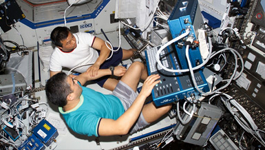
A portable imaging device currently in development by the National Space Biomedical Research Institute (NSBRI) will produce clear, highly detailed pictures of bone and tissue, helping physicians manage bone health in space and on Earth. The Scanning Confocal Acoustic Diagnostic system, or SCAD, will enable doctors to determine the rate of loss and plan treatment options with the aid of high-quality images, taken noninvasively.
Studies of cosmonauts and astronauts who spent months on space station Mir revealed that space travelers can lose, on average, one-to-two percent of bone mass each month, with the greatest loss in the lower extremities like the femur and hip. The culprit is microgravity, which causes bone loss in critical areas and leaves bones susceptible to fracture upon return to Earth.
Space travelers are not the only demographic concerned with bone loss. According to the National Osteoporosis Foundation, at least 10 million people in the United States suffer from bone loss in the form of osteoporosis.
“Because bone weakening is a potentially serious side-effect of extended spaceflight, we’re developing a high-resolution ultrasound imaging device that can monitor and diagnose bone quantity, density and strength in space,” said Dr. Yi-Xian Qin, associate team leader of NSBRI’s technology development team. “We’re currently in the beginning phases of development, but eventually this technology can aid diagnosis for a number of skeletal disorders.”
The real-time, high-resolution, portable imaging device will use scanned, confocal ultrasound for generating images in regions of interest and identifying problems or risk factors. For flight surgeons on the ground, the SCAD will help to quickly determine the rate of bone loss, severity of injuries and possibilities for recovery.
“It will also provide immediate images of bone and assess both density and stiffness data,” said Qin, associate professor in the department of biomedical engineering at Stony Brook University in New York.
Compared to current ultrasound technology for measuring bone loss, the SCAD system provides image-based bone quality parameters in the region of interest, which can be directly related to the assessment of bone strength. The system can also increase the accuracy for ultrasound and reduce the acoustic noise from soft tissue and critical regions via real-time mapping of the bone. It consists of a computer-controlled miniaturized scanner and data acquisition and analysis software designed by a team composed of Stony Brook scientists, a physician and graduate students. In the future, the system will be designed to include wireless output for easy data transmission. The entire device is lightweight and easy to carry.
Qin sees his project in two phases: first; the development phase focusing on miniaturizing the device and fine-tuning its precision, and the second phase; eventual clinical trial and commercial buy-in. From shuttle flights and missions aboard the ISS to future interplanetary travel, the SCAD device could provide immediate emergency diagnosis for injuries or conditions that might otherwise halt a mission.
“Aside from use inflight, my goal is to make this device available to physicians across disciplines to improve the diagnosis of osteopenia and osteoporosis,” Qin said. “Because such diseases are essentially painless at the initial stages, they are difficult to pinpoint and often diagnosed late.”
The SCAD project is complemented by NSBRI teams looking at other space health concerns including adequate sleep, psychosocial factors, cardiovascular changes, muscle wasting, balance and orientation problems, and radiation exposure. While focusing on space health issues, the Institute will quickly transfer solutions to Earth patients suffering from similar conditions.
The NSBRI, funded by NASA, is a consortium of institutions studying the health risks related to long-duration space flight. The Institute’s research and education projects take place at more than 70 institutions across the United States.


