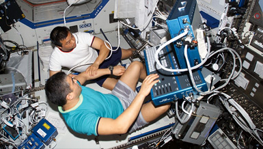
Diagnosing and monitoring health problems in space is hindered by the equipment on board and astronauts’ moderate level of training. Dr. James Thomas hopes to expand the use of a three-dimensional ultrasound in space for clinical use and for research. Medical diagnosis in space could be simplified by producing 3-D images of the body before a space flight, providing a basis of comparison for images taken during a mission.
Overview
Diagnostic Three-Dimensional Ultrasonography: Development of Novel Compression, Segmentation and Registration Techniques for Manned Spaceflight Applications
Principal Investigator:
James D. Thomas, M.D.
Organization:
The Cleveland Clinic Foundation
Technical Summary
Three-dimensional ultrasound has the advantage of acquiring a large anatomic volume from a single ultrasonic window, and thus may be less dependent upon the expertise of the examiner. Furthermore, this large volume may contain sufficient anatomic landmarks to allow unambiguous registration with previously obtained three-dimension data from either ultrasound or other modalities such as magnetic resonance imaging (MRI) or computed tomography (CT). One could thus envision a system by which whole organs or even the entire body would be imaged in three-dimensions prior to launch - data that could be used to compare with subsequently obtained three-dimensional data sets using inflight ultrasonography.
The overall purpose of this grant is therefore to perform ground-based research, development and validation aimed at optimizing diagnostic ultrasound in manned space flight with the following general hypothesis:
Unifying hypothesis: Serial, three-dimensional ultrasound examinations will enhance diagnostic capabilities in manned space flight.
The technical aspects of this program will be pursued with the following specific aims:
- Optimize the acquisition methods for three-dimensional sonography, utilizing reconstruction and real-time techniques.
- Develop techniques for registering anatomical images from two- and three-dimensional ultrasound with those obtained from prior ultrasound examination and from magnetic resonance and computed-tomographic imaging, considered gold standards for noninvasive anatomical imaging.
- Develop tools for abstracting, in an automated fashion, anatomical changes from serial three-dimension and two-dimension ultrasound studies.
- Develop algorithms for the optimal compression of three-dimensional ultrasound images and refine current two-dimensional compression algorithms.
- Assess the ability of novice examiners to obtain three-dimensional sonographic data sets following minimal training.
These objectives will be pursued using data from a variety of in vitro, animal and clinical models. In particular, we will take advantage of a well-established collaboration with the National Institutes of Health, which permits highly sophisticated chronic animal models to be examined with a minimum of additional resources. Although the tools developed here should be applicable to any organ of the body, we will focus our efforts on the kidneys and the heart.
At the conclusion of this project, we anticipate delivering to NSBRI and its Smart Medical System Team a set of algorithms and software for the non-rigid morphological registration and comparison of serial two- and three-dimensional ultrasound data sets and validated algorithms for optimal compression of four-dimensional ultrasound data. In addition to these technical deliverables, our validation work on nephrolithiasis will provide important diagnostic clues for assessing this condition in manned space flight. Similarly, the work on cardiac mass regression following unloading will be invaluable to the NASA research and medical operations community in assessing the impact of long-term space flight on cardiac atrophy and utility of prophylactic countermeasures.


