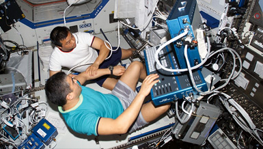
Potential serious risks to astronauts on long missions include reduction in cardiac performance, cardiac dysrhythmias and orthostatic intolerance. As a countermeasure to the possibility that undiagnosed cardiovascular disease could manifest during long missions, Dr. James D. Thomas is working to improve sensitivity for detecting early changes in cardiac function during exercise, exploring echocardiography (ultrasound imaging) as a reliable, portable, noninvasive method to detect and quantify changes that the body undergoes in space, and is improving medical diagnosis in space by producing 3-D images of the body before a flight to provide a basis of comparison for images taken during a mission. In addition, he is continuing his study of the changes that occur in patients’ hearts after aortic valve replacement to determine factors responsible for this loss of muscle in space.
Overview
Echocardiographic Assessment of Cardiovascular Adaptation and Countermeasures in Microgravity
Principal Investigator:
James D. Thomas, M.D.
Organization:
The Cleveland Clinic
Technical Summary
Specific Aims
- Extension of work to calculate two-dimensional myocardial strain, improving sensitivity for detecting preclinical alterations in cardiac function.
- Since early cardiac disease is usually manifest initially during exercise stress, we will develop and validate the tools to apply two-dimensional strain in graded exercise to detect myocardial dysfunction in its earliest phases, allowing both diagnostic capabilities and a means of judging exercise as a countermeasure.
- Continue our ongoing study of the magnitude and predictors of left ventricle mass regression following acute volume and pressure unloading as a ground-based analog for manned spaceflight. This work will continue to focus on patients undergoing aortic valve surgery but will exploit recent knowledge of the roles of cytokines and integrins involved in cardiac hypertrophy and regression as well as emerging technologies such as gene chip analysis.
- Develop, in collaboration with fundamental physics scientists from NASA Glenn Research Center, a sophisticated fluid-structure model of the left ventricle constrained by the pericardium to investigate the impact that microgravity has on unloading the heart by a removal of pericardial constraint.
This work closely focused on risks identified by NASAs Human Research Program. This project will enhance assessment of cardiac function during long-duration missions and potentially suggest cytokine promoters or signal transduction pathways that could be targeted for cardiac atrophy countermeasures.
Earth Applications
The three-dimensional (3D) fluid-structure model of the left ventricle will also have an extensive application in Earth-based research cardiology allowing investigators to alter fundamental inputs for myocardial function and assess the effects on ventricular performance.
Wireless telemedicine systems for ultrasound enable transfer of ultrasound data within the hospital and remotely to workstations connected to our network.
We have continued to investigate 3D ultrasound capabilities. Building on our experience with the Volumetrics system, we have begun to use much improved acquisition devices (Philips ie33 and GE Medical Vivid 7) to obtain 3D examinations in a wide variety of cardiac pathologies.
We have worked on the registration of computed tomography and ultrasound data for improved understanding of both valvular and ventricular function. We are investigating prosthetic valve motion using both modalities to see if 3D ultrasound is able to noninvasively assess function. We are also working on the registration of 3D ultrasound data with nuclear medicine images for assessment of cardiac perfusion.


