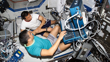
One of the hazards space explorers face is radiation exposure from cosmic and solar sources, which can cause a number of health problems for astronauts. One possible side effect of radiation exposure is the weakening of bones. Since astronauts are also susceptible to bone loss due to reduced gravity in space, the exposure to radiation may compound the risk. NSBRI Postdoctoral Fellow Dr. Jeffrey S. Willey is conducting a research project to better understand how the combination of radiation-exposure and a modeled reduced-gravity environment influence bone quantity and quality. The results will be used in the pursuit of countermeasures to bone loss during space exploration missions and the reduction of the risk of accelerated bone loss leading to osteoporosis and mission-critical fractures in astronauts.
Overview
Radiation Effects on Bone Tissue and Cells in Reduced Gravity (Postdoctoral Fellowship)
Principal Investigator:
Jeffrey S. Willey, Ph.D.
Organization:
Clemson University
Technical Summary
Our intent is to enhance the understanding of the effects of radiation on bone health, particularly within a reduced-gravity environment. Our ultimate goal is to develop appropriate countermeasures for bone loss in astronauts during exploratory missions. The specific aims will analyze the effects of modeled space radiation using scenarios applicable for lunar outpost missions.
Specific Aims
- Examine the response of bone to a combination of spaceflight-associated challenges, including radiation exposure together with reduced limb bone loading.
Approach
Skeletally mature female C57BL/6 mice, a strain sensitive to disuse-associated osteoporosis, were exposed to a 1 Gy dose or protons at Loma Linda Medical Center followed by a 4-week period of hindlimb suspension.Main Findings
The combination of radiation and hindlimb suspension resulted in greater bone loss and deterioration of trabecular microarchitecture than the two challenges individually. Although both skeletal challenges induced atrophy, bone loss due to suspension alone was substantially greater than irradiation alone.While reduced loading on the bone combined with microarchitecture resulted in excessive bone loss relative to either one challenge independently, the causal mechanisms for the stimuli may be independent. A thorough histological investigation was undertaken. Throughout the study, osteoclasts appeared to be more active as an early response in both irradiated groups (irradiated only and irradiated + suspension) than non-irradiated groups.
- Examine the mechanistic causes of changes in bone at the cellular and tissue level following the individual and combined effects of radiation and unloading on mouse hindlimbs.
Approach
Year One: Thirteen-week old C57BL/6 mice received whole body radiation with 2 Gy X-rays. X-rays were chosen for these initial studies, as the relative biological effect of X-rays and protons are similar, and bone loss from a 1 Gy dose has been shown to be similar (-13-15 percent) for both types at one month (preliminary data).Year Two: In an attempt to expand this, NSBRI funded developing technology to develop countermeasures for spaceflight and clinical applications. Sixteen-week old mice were exposed to a 2 Gy dose of X-rays. Some irradiated animals were given a commercially available osteoporosis medication (risedronate) to try to prevent any changes. Bone was examined at week 1, 2 and 3.
To apply this technology to clinical applications, the right hindlimbs of 20-week old Sprague-Dawley rats received a dose of radiation that modeled what the human hip receives during cancer radiotherapy. Animals were sacrificed at 2, 4 and 6 weeks to identify if modeled cancer therapy could induce bone loss. An antiresorptive agent (a commercially available bisphosphonate, risedronate) was applied to try to prevent these changes.
Main Findings
Radiation appears to increase osteoclast number and activity as an early, acute response to radiation leading to early atrophy. Circulating markers of bone resorption are increased by 24 hours of exposure, with significantly increased osteoclast numbers present along trabeculae by three days. Trabecular bone loss is significant and substantial by day seven after exposure and occurs at multiple skeletal sites (proximal tibia; distal femur; fifth lumbar vertebrae). Bone quantity remains lower than control at day 14 and 21.Low-dose irradiation of mice resulted in substantial deterioration of bone one week after exposure. These changes occurred as a result of increased osteoclast activity and active bone resorption. An agent that blocks osteoclast activity (risedronate) completely prevented these changes. Modeling cancer radiotherapy resulted in severe deterioration of trabecular bone in rats at all time points investigated. Applying risedronate completely prevented deterioration of bone early after modeled cancer radiotherapy.
While Year 2 of funding was largely devoted to the development of a therapy for radiation-induced bone loss in both the spaceflight and clinical environment, identification of the causal mechanisms for bone loss must be identified. We have clearly identified that radiation somehow turns on osteoclasts, and that this stimulation of active bone resorption (as opposed to suppressed bone formation) leads to bone loss. Our lab is identifying that markers of inflammation may be turning on these osteoclasts early after exposure. These include tumor necrosis factor-alpha (TNFa) and interleukin-1 (IL-1) activity. Therefore, for the coming year, we will investigate the role of these cytokines in radiation-induced bone loss. We will use a mutant mouse strain (reduced-inflammatory strain) developed in the lab of Dr. Albert Fornace, Jr., (Georgetown University) in collaboration with his NSBRI Postdoctoral Fellow, Dr. Daniella Trani. These mice will be exposed to a series of particle radiation exposures (both protons and iron nuclei; at low doses) at Brookhaven National Laboratory to identify if reducing inflammation can prevent bone loss.
Additionally, we will more appropriately model the skeletal effects of radiation during realistic spaceflight scenarios by irradiating mice with 0, 1 or 2 G ray doses of protons at a prolonged dose rate, modeling the maximum dose rate at the peak of a solar flare.
Earth Applications
Our findings suggest that:
- Radiation increases osteoclast activity and number in the first week after exposure, leading to substantial loss of bone;
- The bone loss that occurs during the first week and any other evidence of deterioration can be prevented by applying a bisphosphonate as an antiresorptive countermeasure; and
- These observations are true in both mice (after low-dose photon exposures) and rats (after receiving targeted and modeled cancer radiation therapy).
Radiation-induced bone damage is a concern for radiation oncologists. Improvements in cancer treatment and diagnosis have led to increased long-term cancer survivorship. For example, of the more than 200,000 men who are diagnosed with prostate cancer each year, the 10-year survival rate approaches 90 percent. However, this growing population of long-term survivors are at risk of developing deleterious effects in normal tissues resulting from the use of therapeutic radiation. Skeletal complications, particularly fractures, are a recognized late radiation-induced effect. Fractures of the hip, ribs, clavicle and humerus have been documented in patients receiving targeted radiotherapy for pelvic and breast tumors. These bones absorb radiation during cancer treatment. The increased incidence of hip (particularly the femoral neck) fractures in postmenopausal women after receiving pelvic cancer treatment is substantial and can have severe negative impacts on a patients mobility, independence and survival.
More post-menopausal women, who fracture a hip, will die within a year of the fracture than will die as a result of breast cancer. Therefore, preventing hip fractures due to radiation therapy (as well as vertebral breaks) are important in maintaining a patients quality of life and health.
Work from this project has identified that by preventing osteoclast activity after radiation exposure, bone loss can be prevented. This work has helped lead to the development of several clinical trials within oncology departments in California and North Carolina. If these observations translate into the clinic, it is possible that the notion of preventing bone deterioration resulting from cancer therapy by targeting the osteoclast can improve patient quality of care.


