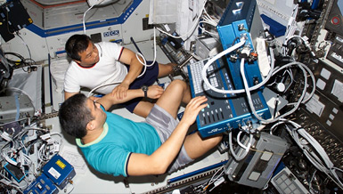
Muscle strength and bone mineral density dramatically decrease in space, but the rate of recovery of muscle versus that of bone has not been well defined yet. However, muscle recovers much faster than bones do which creates an imbalance in strength and increases the risk for fractures. Dr. Susan Bloomfield is investigating the rate and completeness of recovery of muscle strength and bone mineral density and will test various mechanical and pharmaceutical countermeasures that might enhance muscle and/or bone recovery.
Overview
Bone and Muscle Recovery from Simulated Microgravity
Principal Investigator:
Susan A. Bloomfield, Ph.D.
Organization:
Texas A&M University
Technical Summary
Our objectives were to use the well-validated model of the hindlimb unloaded (HU) rat to perform more invasive studies than is feasible in humans to determine the time course of recovery of functional properties in a muscle-bone pair after a period of simulated microgravity, and to test the effectiveness of mechanical and pharmacological interventions in promoting return of bone strength without exacerbating a muscle-bone strength mismatch. The time course studies comprehensively describe the recovery of muscle and bone following 28 days of HU, with in-vivo measures of bone density and geometry and of muscle functional properties. Ex-vivo analyses of bone marrow osteoprogenitor cells and histomorphometric analyses and mechanical testing of cortical and cancellous bone sites were completed.
Key findings include: functional properties of the plantarflexor muscles recovered completely within 14 days of return to normal weight-bearing; normal weight-bearing alone stimulated no recovery of volumetric bone mineral density (vBMD) at the proximal tibia until between 56 and 84 days of recovery, resulting in a significant mismatch between the two tissues strength between 14 and 56 days of recovery; and cell culture studies of marrow from femurs of rats recovering from HU suggest a decline in mature osteoblast activity in spite of greater numbers of bone marrow osteoprogenitor cells.
Future research should better define the clinical relevance of this mismatch between functional properties of muscle-bone pairs during recovery from prolonged disuse, with a particular emphasis on the risk of avulsion fracture. Two interventions were tested during a 28-day recovery period from HU for their ability to stimulate a more rapid recovery of bone with the goal of minimizing the mismatch between bone and muscle strength. Two groups of rats were implanted with tibial nerve cuffs so that stimulated isometric contractions of the plantar flexors could be performed during recovery, mimicking high-intensity resistance training. The high-force muscle stimulation protocol during recovery was only partially effective in enhancing recovery of total and cancellous VBMD at the proximal tibia vs those rats with nerve cuffs that received no training. There was a trend (p =.07) for peak isometric torque of the posterior crural muscles to be greater after 28 days of muscle stimulation as compared to untreated rats.
Downregulation of muscle mass and strength in all nerve-cuff implanted rats complicates interpretation of some of these data. On the other hand, our intervention with an FDA-approved osteoporosis treatment, intermittent PTH, effectively stimulated rapid increases in bone mass at the proximal tibia during recovery, at a rate nearly equal to that for muscle strength. Micro-CT studies documented higher cancellous bone volume in the proximal tibia of PTH-treated rats versus controls, accounted for by higher trabecular thickness and number, and reduced trabecular spacing. Material properties of the proximal tibia cancellous compartment exhibited parallel improvements. The mechanism for these improvements stems from the stimulation of osteoblast activity by PTH. Bone formation rates were increased on cancellous and cortical bone surfaces at proximal and mid-shaft tibia. Hence, both cortical and cancellous bone compartments can respond to the anabolic effects of intermittent PTH treatment after a prolonged period of mechanical unloading.
Because several parameters of bone mass and volume were increased well beyond baseline values after 28 days of PTH treatment (using 80 g/kg BW/d), it is likely that a much smaller dose of PTH would be effective in hastening bone recovery from HU and post-spaceflight. Further work should clarify how low a dose would be effective in stimulating this more rapid return of bone mass, and also whether these gains in bone mass and strength can be retained after short-term PTH treatment is discontinued. Such maintenance may well require the alternative anabolic stimulus of regular exercise.
Earth Applications
Despite evidence in previous studies for depressed osteoblast function after disuse, it appears that, once weight-bearing is resumed, cancellous bone is highly responsive to the anabolic stimulus provided by intermittent PTH. Short-term treatment with intermittent PTH may prove beneficial after prolonged bed rest or unavoidable limb immobilization in accelerating recovery of cancellous VBMD and architecture and restoring normal BFR in cortical bone. Careful dose-response studies should be performed to optimize the dose of intermittent PTH, as the dose used with this small animal model more than replaced the bone lost. Hence, much lower doses of intermittent PTH may be effective. Further research will be needed to define whether PTH-induced gain in bone can be effectively maintained once PTH treatments are withdrawn; it is likely that a continued anabolic stimulus, such as regular weight-bearing exercise, will be required.


