Research

Bone

  • Current Research
  • Previous Research

Overview

Decreased integrin β1 expression of Bone marrow stromal cells (BMSC) isolated from hindlimb unloaded rats is restored with time in culture. BMSCs isolated from normally walking (control) and hind-limb unloaded rats were cultured, and cell lysates were subjected to immunoblotting. Integrin beta1 expression is decreased in cells from unloaded rats compared to control ones after seven days in culture. However, integrin beta1 expression is restored by days 14 and 21. These results are consistent with RNA expression of beta 1 and other subunits of integrin. Image courtesy of Takuo Kubota, M.D., Ph.D. Click here for larger image.

Interaction Between IGF-1 and Integrin Signaling in the Response of Bone to Mechanical Load (Postdoctoral Fellowship)

Interaction Between IGF-1 and Integrin Signaling in the Response of Bone to Mechanical Load (Postdoctoral Fellowship)

The working model for this project is that integrins activated by mechanical loading are necessary for insulin-like growth factor type 1 receptor (IGF-1R) activation induced by IGF-1. In skeletal unloading, impaired integrin expression leads to a decrease in IGF-1R activation, resulting in a reduction in bone formation. In contrast, loading maintains integrin expression and enhances IGF-1R activation, leading to an increase in bone formation. Integrin composed of α and β subunit is positioned to interact with extracellular matrix and transduce mechanical forces into biochemical signals. Image courtesy of Takuo Kubota, M.D., Ph.D. Click here for larger image.

Principal Investigator:
Takuo Kubota, M.D., Ph.D.

Organization:
University of California, San Francisco

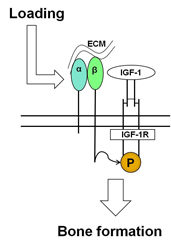
Bone loss is a serious health risk of living in the reduced gravity of space for long periods of time. In space, bones no longer experience the stimulation, or loading, that occurs during activities such as walking and running in normal gravity. Scientists do not fully understand the mechanisms that cause this bone loss. Research indicates that insulin-like growth factor type 1 (IGF-1), a hormone that plays a role in childhood growth and in tissue building in adults, is ivolved in bone formation after load-bearing activities such as walking. When weight-bearing activity is removed, IGF-1 fails to increase bone formation.

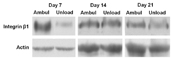
Dr. Kubota’s project seeks to understand what role the IGF-1 receptor (IGF-1R) and beta 1 and beta 3 integrins (which help cells attach to tissues) play in the formation of bone following load-bearing activity. The study will examine changes in mice lacking IGF-1R and beta 1 and 3 integrins when the mice are placed in conditions simulating reduced gravity followed by normal gravity. The project seeks to learn more about the role of IGF-1 and beta 1 and 3 integrins in spaceflight-induced bone loss.

NASA Taskbook Entry


Technical Summary

Bone loss resulting from the lack of mechanical stimulation in the weightlessness of spaceflight is a significant concern for prolonged space exploration missions. However, the mechanisms of bone loss in microgravity have not been fully understood. Evidence supports a role for insulin-like growth factor type 1 (IGF-1) signaling in mediating skeletal response to mechanical load. Skeletal unloading by hindlimb elevation, a ground-based model for the unloading of spaceflight, causes bone loss due to reduced bone formation. IGF-1 is a potent growth factor in bone and is produced in osteocytes and mature osteoblasts after mechanical load. This IGF-1 may produce new osteoblasts and differentiate them into mature osteoblasts to form new bone. Furthermore, skeletal unloading leads to resistance to the anabolic effect of IGF-1.

IGF-1 fails to increase bone formation and osteoblast proliferation in the unloaded bone. This failure to respond to IGF-1 is due to a failure of IGF-1 to stimulate the phosphorylation of the IGF-1 receptor (IGF-1R), although IGF-1R levels and binding of IGF-1 to its receptor are not changed. Skeletal unloading decreases integrin expression in osteoblasts, and this impairment of expression is restored by reloading the bones or the osteoblasts in parallel with the recovery of IGF-1 responsiveness. Knockdown of beta 1 and beta 3 integrins blocks the ability of IGF-1 to activate its receptor and inhibits its activation by mechanical loading.

This project will use axial cyclic loading of the tibia as well as reloading following skeletal unloading in mice lacking IGF-1R or beta 1 and beta 3 integrins to determine the importance of these molecules in the response of bone to mechanical load. We will also use pulsatile fluid flow, a mechanical loading modality in vitro, to determine the molecular mechanism by which integrin signaling by mechanical load enhances IGF-1 signaling. If this hypothesis proves to be true, efforts to maintain the integrin signaling and IGF-1 signaling will be sought to prevent bone loss associated with skeletal unloading.

Key Findings
Mice lacking the IGF-1R in mature osteoblasts and osteocytes were subjected to skeletal unloading using hindlimb unloading and reloading either after unloading or with cyclic axial loading of the right tibia during hindlimb unloading. μCT analysis showed that the response of bone volume fraction (BV/TV) of the trabecular bone in the distal femur to unloading and reloading is similar in IGF-1R knockout (KO) mice to that in control mice at baseline and after two weeks of unloading and two weeks of reloading. Moreover, the response of the trabecular bone and cortical bone in the tibia to cyclic loading was comparable in KO mice to control mice. To further investigate response of the KO mice to reloading, bone histomorphometric analysis was performed. Bone formation rate, mineral apposition rate and mineralizing surface were markedly reduced in the periosteal cortical bone of the tibia of the reloaded KO mice after unloading compared to those in the control mice.

Impact of Key Findings
The above findings indicate that IGF-1R plays a significant role in response to reloading after unloading, at least in the cortical bone. Bone histomorphometric analysis in the trabecular bone is being performed to elucidate the underlying cellular mechanisms. We postulate that bone resorption is also reduced during the reloading after the unloading perhaps secondary to the reduction in bone formation.

Proposed Research Plan
We will test mice deleted for integrin beta 1 and beta 3 from mature osteobasts and osteocytes to determine whether the skeletal response to mechanical load and IGF-1 infusion is impaired. We will also knock-down or inhibit selected components of the integrin/IGF-1R complex to determine whether this blocks IGF-1R activation and/or the formation of the integrin/IGF-1R complex during pulsatile fluid flow.

Earth Applications

Bone loss is a serious problem in immobilized people due to injury or surgery on Earth. A reduction in bone mass raises a risk of bone fracture. It takes a long time for those patients to recover bone strength in rehabilitation. The mechanisms of bone gain after weight-bearing activity following long-term bed rest have not been fully elucidated as well as those of bone loss during immobilization. Insulin-like growth factor 1 (IGF-1) signaling plays a vital role in bone remodeling. IGF-1 production is increased in osteocytes and osteoblasts after mechanical load. Skeletal unloading by hindlimb elevation, an animal model for immobilization on Earth as well as a ground-based model for the unloading of spaceflight, causes a decrease in bone mass associated with resistance to the anabolic effect of IGF-1.

We postulated that IGF-1 signaling mediates the skeletal response to unloading and reloading. To determine whether the IGF-1 receptor (IGF-1R) in mature osteoblasts and osteocytes has a role in the skeletal response to unloading and reloading, we evaluated mice in which the IGF-1R was deleted with osteocalcin-driven cre recombinase. These mice were subjected to skeletal unloading using hindlimb unloading and reloading after unloading. We found that IGF-1 signaling was critical for bone formation after reloading following skeletal unloading. Bone formation rate, mineral apposition rate and mineralizing surface were decreased in the periosteal cortical bone of IGF-1R deficient mice compared to control mice. These results suggest that IGF-1 signaling is necessary for promoting bone formation after weight-bearing activity following immobilization on Earth and that efforts to maintain and potentiate the IGF-1 signaling will be sought to enhance bone formation following long-term bed rest.

This project's funding ended in 2011