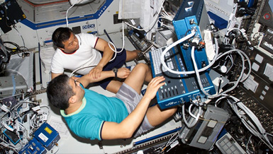
Exposure to microgravity during long-duration space flight can decrease bone and muscle mass and increase the risk of fractures. Dr. Harry K. Charles, Jr., is developing a compact, lightweight instrument to accurately and precisely determine bone density and geometry during space travel. Charles’ device will help scientists evaluate the risk of bone fracture and determine the effectiveness of countermeasures. On Earth, Charles’ instruments will allow doctors to better monitor and treat bone loss associated with osteoporosis and lack of mobility.
Overview
AMPDXA Scanner for Precision Bone and Muscle Loss Measurements During Long-Term Spaceflight
Principal Investigator:
Harry K. Charles, Jr., Ph.D
Organization:
The Johns Hopkins University Applied Physics Laboratory
Technical Summary
The AMPDXA project is a joint effort between the NSBRI's Technology Development Team, the Bone Demineralization/Calcium Metabolism Team, and the Muscle Alterations and Atrophy Team. Its goal is to provide the high precision monitoring system necessary to fully assess both the deleterious effects of weightlessness on the bones and muscles and the effectiveness of any countermeasures. We believe that any pharmacological or exercise-related countermeasures used by astronauts to mitigate microgravity effects will require efficient and timely monitoring. Moreover, the monitoring device must be capable of being used by astronauts during spaceflight so that feedback can be dynamically employed to regulate countermeasure doses. The system design will be such that intelligent, but not necessarily medically trained, personnel will be able to create scans that will provide all of the accuracy and precision necessary. Readouts and displays for the AMPDXA instrumentation will be specifically designed to provide useful (real-time) feedback information to both the astronauts and the ground-based physician monitoring team (as permitted by the mission dynamics).
Current bone and muscle mass measurements (via conventional DXA or ultrasound) are regional averages that obscure structural details. Since the mechanical consequences of lost bone and muscle are reflected in the structure, an absolute determination of skeletal mechanical competence is needed to supplement the loss measurements. Engineering properties of the bones can be derived from DXA-generated BMD data. Our method derives geometrical measurements from the BMD images. From such images, we extract BMD profiles at important skeletal locations (e.g., proximal shaft and femoral neck). Key properties measured and derived from these profiles include the BMD, the subperiosteal width, the section modulus (related to strength), and the cortical dimensions.
Under the original proposal effort, FY 1998-2000, the AMPDXA project made significant progress in several key areas: (1) instrument development, (2) algorithm development for BMD image extraction and structural analysis, and (3) bone reconstruction and modeling techniques. During the FY 1998-2001 period, both a full-sized (one-meter source-to-detector distance) Laboratory Test Bed and a system for human testing were constructed. This system was initially called the Clinical Test System in previous reports, but is now called the Human Test Bed to better reflect the nature of the human testing to be performed on the system. The Laboratory Test Bed was utilized to verify principles and theoretical predictions and demonstrate that the AMPDXA techniques worked and produced results with the expected precision. The Human Test Bed has even greater precision.
The Human Test Bed incorporates high-precision, rotational and translational stages to provide the scanning capability to carry out qualification tests on human subjects. Since the Human Test Bed is designed to operate only on Earth, the table, gantry, and associated equipment were not built to the size and mass requirements of an AMPDXA unit for spaceflight. In fact, the unit was built on a used CT scanner. Employing used equipment for some of the structural elements and rotating parts and machinery allowed critical resources to be focused on the information extraction and analysis issues leading to human testing.
The image extraction capability of the AMPDXA is not only the BMD image higher resolution, but also the mass distribution in a projected thickness of a femur slice contains much more structural detail than conventional DXAs. The high frequency content of the BMD spatial projections are reproducible and provide information on the bone's microstructure. Using multiple projections (three or greater) about the bone axis allows structural properties (e.g., bending strength) to be obtained independent of patient position. Initial experimental measurements with different sets of three projections showed that the principal moments of inertia could be determined within three to four percent. Additional projections (above three) reduce this number further. Our original experimental system also had some known non-linearities, which have since been removed, and our error in the three-projection estimation of moments is less than one percent.
For the 2002 period, we are focused on resolving certain key issues about the AMPDXA and then successfully using the AMPDXA for human testing. These key issues include: (1) unequivocal demonstration that multiple projection technology improves BMD accuracy and collects structural details, (2) the structural details can be converted into bone reconstruction models that preserve mechanical behavior, (3) the reconstruction models can be utilized to predict risk of fracture, (4) soft tissue can effectively be distinguished from bone and decomposed into fat and muscle, (5) data can be collected reliably and repeatedly on human subjects using the Human Test Bed, and (6) the Human Test Bed can be utilized in research studies on bone and muscle loss.
A dual monitor computer system currently operates the AMPDXA as well as records and displays image maps (bone mineral density, muscle, etc.) at near real-time speeds. The main screen presents two views of the BMD images of a human hip collected from a live human test subject. The major accomplishments during the period include reverification and calibration of the Human Test Bed after the move, improving the AMPDXA operational software (providing full documentation and configuration control), refining the image extraction algorithms, demonstrating the Human Test Bed's accuracy and repeatability, and human imaging. Approval for our human testing protocol has been granted by the Institutional Review Board at the Johns Hopkins Medical Institutions.
The AMPDXA project has many implications for future research and development. The AMPDXA, as described above, has direct application to risk reduction in NASA's Critical Research Path. The AMPDXA is capable of real-time monitoring of bone and muscle loss at extremely high precision. Since the results are patient-specific and not tied to volumetric averages and statistical norms, the AMPDXA is a very useful tool for monitoring the effectiveness of countermeasures as well as determining risk of fracture under various loading conditions and activity scenarios. The AMPDXA also appears to be a natural adjunct to Earth-bound research on the effects of aging and disuse on bone integrity. It could also be used as a routine screening tool for osteoporosis and as a monitoring instrument for osteoporosis drug therapy.


