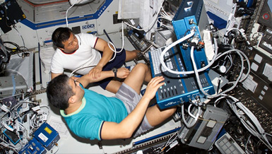
The loss of bone density resulting from the weightless environment of space is a major health issue for astronauts during long duration spaceflight. Dr. Harry K. Charles, Jr. and his colleagues – both at the Johns Hopkins University Applied Physics Laboratory and the School of Medicine – are building a ground-based clinical system using Advanced Multiple Projection Dual-Energy X-ray Absorptiometry (AMPDXA) techniques to measure bone and muscle loss and determine changes in bone structure. Performing high precision scans on astronauts before and after spaceflights with the AMPDXA will enable flight surgeons to monitor musculoskeletal strength and to determine the effectiveness of countermeasures. This system could have a large impact on the bone health of our aging Earth-bound population because both bone research and clinical medicine will benefit from a more definitive method for diagnosing osteoporosis and other bone disorders, monitoring progress and assessing the efficacy of treatments.
Overview
Ground-Based Measurement of Bone Loss in Astronauts Using AMPDXA Ground-Based Clinical System
Principal Investigator:
Harry K. Charles, Jr., Ph.D.
Organization:
The Johns Hopkins University Applied Physics Laboratory
Technical Summary
The goal of the AMPDXA Ground-Based Clinical System (AMPDXA-GCS) was to produce an operational instrument that could be transferred to NASA Johnson Space Center for use in the pre-and post-flight bone mineral density and bone structure measurements on astronauts.
Original Aims
- Design, fabrication and testing of AMPDXA-GCS.
- Validate the utility, radiation safety and method of operation of the AMPDXA-GCS configuration.
- Address key technical challenges necessary to migrate the AMPDXA-GCS from ground-based studies to spaceflight.
- Transfer AMPDXA-GCS technology into commercial clinical uses.
An imaging system with AMPDXA-GCS qualities in space would be an unrivaled tool for rapid, high-precision testing of the effects of countermeasures on the musculoskeletal system. Thus, AMPDXA-GCS technology is highly likely to result in new tests for countermeasures efficacy, and it promises to make a significant contribution to risk reduction.
AMPDXA-GCS monitoring techniques could also benefit millions of people suffering from osteoporosis and other bone disorders. Research and clinical medicine will both benefit from increased ability to more precisely monitor bone health and the efficacy of treatments. Plus, the design principles will allow the development of a compact, less expensive version for limb imaging that could be conveniently used in a variety of remote settings such as rural clinics and nursing homes.
The AMPDXA-GCS builds on the successes demonstrated in previous NSBRI-funded collaborations between the JHU APL and the JHU SOM which resulted in an AMPDXA Laboratory Test Bed and a Human Test Bed. Since the AMPDXA-GSC will operate on slightly different principles (three X-ray beams at fixed angles) than the previous system design, it was necessary to demonstrate the new concept using a test system: the AMPDXA-GCS Test Bed (ATB). The ATB was constructed and initial operating tests verified the new single scan with three fixed-beams principles. With the realization that due to lack of funding the AMPDXA-GCS would not be built, the investigators turned their attention and remaining funding (with NSBRI permission) toward demonstration of the AMPDXA-GCS principles on a commercial duel energy x-ray absorptiometry (DEXA) scanner with the goal of commercializing the new technology invented under NSBRI funding.
To this end, the JHU team began working with the latest Hologic DEXA scanner and was able to modify text-based scan protocols available in the system to allow multiple projection imaging. Multiple projection images were obtained from a sample bone at a constant distance by properly rotating the C arm and translating the patient table. We were then able to convert the Hologic data files into x-ray attenuation files using a visual basic program. Software operations on these files resulted in bone mineral decomposition files. Several scans were made on phantoms and various bone samples such as a pig femur. At this point, we were able to use our previous software to calculate bone cross sectional properties and compare the results to previous scans of the same bone using computed tomography (CT). A good comparison resulted between the Hologic-derived properties, CT scan (used as reference) and our previous AMPDXA scanner. This proved that a commercial system could be modified to provide improved bone analysis embracing AMPDXA-GCS principles. Because of a lack of source code, the scanning protocols developed were slow and would need to be improved before human testing could begin.
Earth Applications
Since the AMPDXA-GCS measures structure to a high precision and does an excellent job at imaging not only bone but also man-made prosthetic implants, the AMPDXA-GCS could be used by orthopedic surgeons to study the life progression of implants (e.g., loosening, bone regression, etc.). Given its low radiation dose and ability to automatically determine dimensions from the collected radiographic data, the AMPDXA-GCS may prove valuable in other orthopedic applications as well.
For example, it is possible to predict stress fractures from bone mineral density measurements. Stress fractures in military recruits range from one to seven percent, depending upon gender and branch of service. An untold number of athletes and other individuals also suffer stress fractures annually as a result of intense repetitive exercise. Such exercise or other similar activity can tire muscles, so that they are no longer able to absorb the shock from these activities. As a result, tiny hairline cracks known as stress fractures develop in the bone. The best treatment for stress fractures is rest for six-to-eight weeks. Such rest periods would prevent a professional athlete or a soldier from performing his/her job. Thus, the ability to identify persons susceptible to stress fractures would enable a different training regimen for these persons in order to prevent the onset of a stress fracture.
Commercially, if cost projections can be realized, there is a significant market for population screening and treatment monitoring of osteoporosis. Based on the AMPDXA-GCS, an easy-to-use, relatively compact instrument could be developed for a small clinic or doctor practice. The unit could be either horizontal like the GCS or vertical (patient stands). Concepts for a vertical unit have been developed. The market for such a commercial AMPDXA could range from several hundred to several thousands of units, depending upon final cost. If the unit could be structured to be lightweight and relatively transportable, it would have important implications for use as a tool in nursing homes, clinics and doctors offices. Several key members of the NSBRIs Musculoskeletal Alterations Team have already indicated that a precision AMPDXA would greatly assist their work in bone loss studies, especially in bed-rest studies.
AMPDXA commercialization efforts are being pursued by both the APL Technology Transfer Office and the Technology Transfer Office at the Johns Hopkins Medical Institutions. Commercialization of the AMPDXA software for use with conventional DXA data may provide the stepping stone for the commercialization of the AMPDXA hardware.


