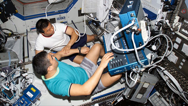
Doctors currently use magnetic resonance imaging (MRI) instruments to depict internal body structures, and Dr. Isaac N. Bankman and his team are investigating the feasibility of a higher-resolution, lighter, MRI system for space. The system would enhance the study of the physiological alterations and aid the development, verification and maintenance of countermeasures.
Overview
Development of a Space Qualifiable MRI System
Principal Investigator:
Isaac N. Bankman, Ph.D.
Organization:
Johns Hopkins University Applied Physics Laboratory
Technical Summary
Mice and small rat models are useful surrogates to carry out in-orbit physiological studies. Measuring alterations in the limbs of astronauts, especially the lower limbs, will provide partial confirmation of the effectiveness of proposed countermeasures and the utility of Earth-based animal models. Inflight MR imaging of mice and rats will especially benefit the countermeasure developments of several of the NSBRI research teams.
The proposed concept is based on traditional MRI principles and uses advanced technology and advanced engineering techniques to reduce mass and power to acceptable levels. The system consists of a one to one and a half Tesla cryogen-free, high-temperature superconducting magnet subsystem and advanced electronics that will have magnetic field inhomogeneities less than or equal to eight ppm over a spherical imaging volume of ten cm diameter and less than or equal to ten ppm out to 15 cm diameter. The magnet cryocooler subsystem will be designed using high temperature superconducting materials to significantly reduce the mass and power of the cryocooler.
The highest resolution mode gives a resolution of 117 microns for small animals over a spherical imaging volume of six cm diameter and a resolution of 352 microns for human limbs over a spherical imaging volume of 18 cm diameter. The standard resolution mode will provide a resolution of 234 microns and 703 microns, respectively. The pulse sequence scenarios used will be those traditionally used in MR imaging to achieve images that are proton-density, T1 or T2 weighted, so that a significant amount of structural information will be available. Because of budget limitations, only selected electronics will be reengineered to demonstrate the minimum mass and power that can be achieved.
The team is composed of individuals and organizations with a unique combination of expertise including: MRI systems development at the General Electric Research and Development Center, advanced MRI development and small animal experimentation at the Johns Hopkins School of Medicine, and the development of reliable medical and low-mass, low-power systems for space applications at the Johns Hopkins University Applied Physics Laboratory.


