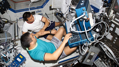
Astronauts who perform spacewalks are at risk for decompression sickness (“the bends”). If decompression sickness occurs, it can cause severe joint pain, coughing, skin irritation, cramps and even paralysis due to nitrogen bubbles in the blood and tissue. Dr. Jay C. Buckey and Creare, Inc. are testing ultrasonic instruments that detect small nitrogen bubbles in both blood and tissue to monitor for and prevent decompression sickness. If successful, these instruments will lead to better understanding and detection of decompression sickness in astronauts as well as deep-sea divers and pilots.
Overview
Improved Bubble Detection for Extravehicular Activity
Principal Investigator:
Jay C. Buckey, Jr., M.D.
Organization:
Dartmouth Hitchcock Medical Center
Technical Summary
Progress
Progress toward establishing the appropriate transducer configurations, electronic settings and instrument enhancements to detect and size bubbles reliably in vivo - This aim has been accomplished. This was done using a stepwise approach. First, experiments were performed in-vivo using agitated saline (the agitated saline contains small bubbles). The transducers of the dual frequency device were aimed into the right ventricle. Agitated saline was injected intravenously while data were collected with the device. These experiments demonstrated that bubbles could be detected as they move through the right ventricle and right atrium. These experiments established the technical knowledge (transducer position relative to anatomical features, equipment settings, etc.) needed to monitor bubbles during subsequent decompression experiments.
In the decompression experiments, the transducers were positioned on the chest wall and the ultrasonic energy was beamed into the right ventricle. The pump frequency (which selects the size of bubble that will be imaged) was increased stepwise from 30 kHz to 180 kHz in 5 kHz increments. At each frequency data were taken with the pump transducer on and off. By comparing the signals returned with the pump on to that with the pump off for each pump frequency, a histogram of bubble sizes could be produced. This work is significant, since the ability to produce bubble size histograms during decompression stress is a new capability that may have both operational and research uses.
Progress toward comparing the new bubble monitoring technique to Doppler, and using it to investigate decompression sickness - This aim has been advanced by comparing the signals obtained with the dual frequency device to a standard clinical ultrasound instrument. Early indications are that the dual frequency device may detect bubbles prior to Doppler, but work in this area is ongoing. A Doppler capability has also been added to the dual frequency device, primarily to assist in aiming the transducers at the ventricle but also as a complementary means to signify the presence of bubbles.
The combination of the Doppler and dual frequency ultrasound is being used to: (a) evaluate the changes in bubble size during the evolution of decompression sickness and (b) evaluate perfluorocarbons as a potential treatment for decompression sickness. The combination of the two bubble detection capabilities into one device provides a versatile instrument for studying decompression sickness.
Progress toward developing the capability to size small bubbles in tissue - This aim has been advanced through a variety of in vitro and in vivo studies. The tissue bubble detection effort has two goals: (a) to evaluate whether very small bubbles (< 30 microns) can be detected in tissue, since decompression sickness theory suggests that small bubbles may exist in tissue normally at ambient pressure and (b) to detect larger bubbles in tissue and in the vasculature that may cause symptoms during decompression sickness. Current efforts have focused on the first goal. Early studies demonstrated signals consistent with bubbles in the thigh of the swine. These signals were found only at particular locations. In the original implementation of the detector, however, the source of the mixing signals could not be determined because of poor spatial resolution. The modified bubble detector now allows for sampling at selected depths, so the mixing signals can be correlated with anatomic structures. This has shown that mixing signals are most likely returned from interfascial planes, i.e. the areas between muscle groups.
Validating tissue bubble detection requires a reliable way to demonstrate that signals detected in tissue originate from bubbles. Current research is focused on developing in-vitro tissue bubble simulators capable of generating prototypical small bubbles to test the tissue bubble detection equipment. Several in-vitro methods are under evaluation, including contrast agent embedded in gelatin, decompressed gelatin, and schemes involving the passage of high-pressure air through very small pipettes.
The tissue bubble detection work is significant since the ability to detect and size bubbles in tissue would be a new and unique capability.
Plan for the Coming Year
In the coming year the plans are to:
- Refine the bubble size histogram capability;
- Use histograms to evaluate bubble sizes during decomrpession stress and during interventions to treat decompression sickness (e.g. administration of perfluorocarbons);
- Advance tissue bubble detection; and
- Pursue human use approval for the device.


