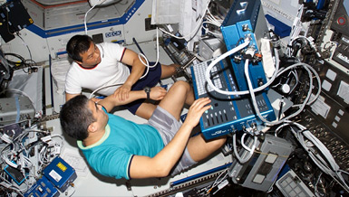
Current imaging devices for the human brain are too heavy and high-powered for space travel. A team headed by Dr. Jeffery Sutton is applying a noninvasive imaging system that is both portable and low-powered to monitor changes in brain activity associated with performance tasks relevant for space missions. The technology, diffuse optical tomography, works by shining light harmlessly through the skull, measuring the light’s reflectance and detecting patterns of brain activity. These patterns are overlaid on three-dimensional models of the brain to help localize where activity occurs. In this way, one can noninvasively monitor the brain at work and investigate, in real time, changes associated with arousal and weightlessness. On Earth, the technology can also be used for brain assessment, diagnosis and monitoring for treatment in various brain disorders, such as stroke.
Overview
Near Infrared Brain Imaging for Space Medicine
Principal Investigator:
Jeffrey P. Sutton, M.D., Ph.D.
Organization:
Baylor College of Medicine
Technical Summary
- Quantitatively assess physiological adaptation (e.g., changes in intracranial pressure and blood flow) associated with microgravity,
- Detect regional brain activity correlated with performance under altered circadian and mental loads,
- Provide remote clinical assessment, and;
- Guide treatment.
In addition to applications for space, the developments of this project have relevance to Earth medical research and care. For example, DOT is now being tested for monitoring stroke progression in patients with acute cerebrovascular accidents. The project brings together scientists, engineers and physicians at the Massachusetts General Hospital/Harvard-MIT Division Health Sciences and Technology, and medical operations personnel at NASA Johnson Space Center (JSC), to work collaboratively on the development and testing of DOT as a space-relevant technology.
The original aims are to:
- Refine current DOT technology to build an instrument with improved spatial and temporal resolution to detect brain activity non-invasively, and in real time, through the intact skull;
- Validate the improved instrument using functional magnetic resonance imaging (fMRI), which is a standard technology, and test DOT as a portable brain imaging device for assessing motor and cognitive activity under normal and sleep deprived conditions in normal human subjects;
- Assess DOT, along with optical coherence tomography (OCT), to non-invasively measure altered intracranial pressure (ICP) in neurological patients, given that altered ICP may occur in the space environment, and;
- Refine a system for automated image interpretation using individualized brain models and computational techniques.
During the third and final year of this award, including supplementary extension, key findings and accomplishments include:
- Continued refinement of DOT instrumentation to validate the technology using fMRI;
- Simultaneous application of DOT and fMRI to detect brain activity differences during SpaceDock task performance (a custom-designed space-relevant visuomotor performance task);
- Simultaneous application of DOT and fMRI to detect brain activity differences associated with sleep deprivation;
- Manuscript completed for the sleep-deprivation application data (2 manuscripts under review);
- Further development of a demonstration system for automated multi-sensor analysis and interpretation (Automated Intelligent Medical System; AIMS), which can use sensor information from multiple astronauts and/or the environment to make medical diagnoses;
- Manuscript completed describing the AIMS computational system and characterization results, and;
- Leveraging of the NSBRI funding to win an R21 award to investigate in greater detail the application of DOT in patients with altered intracranial pressure.
The impact of these findings are that they address the technology requirements set forth in Aim 1, and provide validation for DOT as laid out in the objectives and hypotheses of Aim 2. The previously acquired motor data using simultaneous DOT and fMRI confirm one of the hypotheses contained in Aim 2, namely that DOT and fMRI will be able to detect changes in regional brain activity contralateral to motor movement. The DOT/fMRI findings using the SpaceDock task confirm the hypothesis that the DOT technique can detect frontal cortex differences as a function of mental load in normal and sleep-deprived subjects. The AIMS system provides a general-purpose platform for simultaneously and autonomously monitoring many sensors, and speaks to the issue of individualized, digitized human, anatomical brain models upon which time-derivative functional data are used for interpretation and display in real time (Aim 4).
In the coming year, under the new R21 funding, patient populations with elevated ICP will be investigated in considerable detail. Finally, continued development will take place on the AIMS informatics system towards more automated machine/human interface, applicable to DOT imaging as well as other, analog settings.
Earth Applications
- Functional brain imaging of (or even during) rehabilitation from stroke or brain injury
- Cerebral imaging or monitoring in remote settings (rural health centers, mountainside, etc.)
- Detection of tissue ischemia (esp. muscle, abdominal, brain)
- Pediatric brain imaging/evaluation (particularly for young, highly-animated patients)
- Stroke detection and monitoring
- Breast cancer detection
- Autonomous medical diagnosis in remote (on-location) settings


