Combined Scanning Confocal Ultrasound Diagnostic and Treatment System for Bone Quality Assessment and Fracture Healing
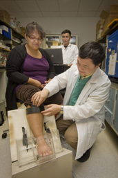
Dr. Yi-Xian Qin (right) and Dr. Jiqi Cheng prepare to use the Scanning Confocal Acoustic Navigation (SCAN) device to collect an ultrasound image of the heel. The new SCAN technology will allow early prediction of bone disorders and guided acceleration of fracture healing and is being developed by NSBRI for use by astronauts during spaceflights. SCAN will also have benefits for health on Earth. (Photo by John Griffin/Stony Brook University). Click here for larger image.
Principal Investigator:
Yi-Xian Qin, Ph.D.
Organization:
Stony Brook University - State University of New York
Astronauts on long-duration spaceflights and the elderly on Earth are more susceptible to fractures due to the loss of bone strength and structure. Early detection and treatment of bone loss will go a long way in preventing fractures and nonunion. The current measurement method, X-ray absorptiometry, is limited in its ability to give a complete picture of bone health. Ultrasound technology – scanning confocal acoustic diagnostic (SCAD) – has shown the potential of providing measurements of bone structure and strength and of guiding effective treatment. Ultrasound has shown potential to accelerate fracture healing.
Dr. Yi-Xian Qin and his colleagues are developing combined diagnostic and treatment ultrasound technology for early prediction of bone disorders and guided acceleration of fracture healing, using SCAD imaging and low-intensity pulse ultrasound.
NASA Taskbook Entry
Using a newly developed noninvasive Scanning Confocal Acoustic Navigation (SCAN) technology, strong correlations between SCAN determined data and bone's structural and strength parameters were observed. Ultrasound has also been shown therapeutic potentials to accelerate fracture healing. The objectives of this study are to develop a combined diagnostic and treatment ultrasound technology for early prediction of bone disorder and guided acceleration of fracture healing, using SCAN imaging and low-intensity pulse ultrasound. The technology will target to the critical skeletal sites, where may be significantly affected by disuse osteopenia and potentially at the risk of fracture.
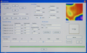
The team is continuing the technology development of a new generation of the SCAN device to access the bone quality at multiple skeletal sites, and use ultrasound to treat the bone fracture in this year. A demo device was shown at the NSBRI five-year review in Houston in January, 2011. A combined mechanical and electrical array scan modality has been initiated, which can complete the SCAN time at the particular skeletal site less in than 2.5 minutes. The new development is capable of generating non-invasive, high-resolution quantitative ultrasound (QUS) attenuation and velocity maps of bone for determining the relationship between ultrasonic specific parameters and bone mineral density (BMD) and bone’s physical properties (i.e., stiffness). Several studies were conducted:
1) Development of electronic array SCAN for bone imaging. The objective for this project is to develop computer-controlled phase delay and sequence of acoustic excitation energy for electronic focusing in the 3-D object. An accelerated continuous scan mode is further designed and built including rapid A/D data acquisition, microprocessor control synchronizing (for scanning, transmit signal, and A/D trigger) and
control algorithm. A high-resolution ultrasound image array with 60x60 (mm2) and 0.5 mm resolution results in scan times of less than 2.5 minutes in the region of interest (ROI).
2) Development of SCAN for multi-site quantitative ultrasound measurement. The team is continue to develop an image based SCAN technology for enhanced diagnostic readings at multiple anatomical sites, e.g., wrist region for distal radius and ulna. A soft contact transducer holder was designed to provide acoustic coupling free of water. The design was validated using 2D SCAN imaging system, and evaluated with its performance at the distal radius. Strong correlation between H2O and gel coupling (R2=0.89), and high repeatability (95%) were observed at the tested site. Results indicated that there was an excellent relationship between ultrasound imaging and microradiographic image. These data demonstrated feasibility of ultrasound imaging in multiple critical skeletal sites, i.e., wrist.
3) Characterization of cortical bone fracture with SCAN and longitudinal acoustic velocity. The objectives of this study were to evaluate the cortical fracture gap size using quantitative ultrasound imaging, and the longitudinal ultrasound velocity in bone to predict the fracture gap size. Strong correlations were observed between ultrasound and X-ray images in fracture size (R2=0.91). High correlation was found between gap size and the longitudinal velocity from 4000 m/s to 3000 m/s for 0-5 mm gaps (R2=0.93). These results suggest that ultrasound is capable to predict bone fracture, and provide useful information for longitudinal assessment of complications, such as non-union fracture, and for evaluating healing.
4) Improving ultrasound sensitivity with phase cancellation method. Phase cancellation in ultrasound due to large receiver size has been proposed as a contributing factor to the inaccuracy of estimating broadband ultrasound attenuation (BUA). In vitro ultrasound measurements were conducted on 54 trabecular bone samples (harvested from sheep femurs) in a confocal configuration with a focused transmitter and synthesized focused receivers of different aperture sizes. Phase sensitive (PS) and phase insensitive (PI) detections were performed. The results indicate that the receiver aperture size used in the confocal configuration with PI detection should at least equal the aperture of the transmitter to capture most of the energy redistributed by the interference and diffraction from the trabecular bone.
5) Mitigation of bone loss using guided therapeutic ultrasound in osteopenia. The objective of this project is to evaluate the capability of guided ultrasound in generating dynamic mechanical signal to inhibit bone loss in an estrogen deficient model of osteopenia. Specimen specific, voxel based, finite element (FE) models (E=18GPa & ν=0.3), were generated using μCT image data and 1% axial compressive strain was simulated using a nonlinear FE solver (ABAQUS). US treatment significantly increased BVF compared to OVX controls for the 100mW/cm2 treated group. This study suggests that there exists a minimum intensity threshold below which US in less effective at maintaining bone’s microstructural and mechanical characteristics. The paper is published in BONE.
6) Acceleration of fracture healing in vivo in a disuse animal model using guided ultrasound. Long duration microgravity not only induces bone loss, but also increases risk of fracture. The aim of this project is to evaluate the potentials of low-intensity guided therapeutic ultrasound on acceleration of fracture healing under delayed union model using hindlimb suspension (HLS) rats. The results have revealed that 6% less bone volume was observed in HLS alone group than the normal fracture (i.e., normal non-HLS condition). Ultrasound treatment on HLS fracture accelerated the healing with significant higher bone volume by 25% more than the HLS alone group. These data imply that guided ultrasound can speed up the healing and mineralization in the fracture callus, while disuse may delay the fracture healing.
Musculoskeletal decay due to a microgravity environment has greatly impacted the United State’s civil space missions and ground operations. Such musculoskeletal complications are also major health problems on Earth (i.e., osteoporosis and the delayed healing of fractures). About 13 to 18 percent of women aged 50 years and older and 3 to 6 percent of men aged 50 years and older have osteoporosis in the U.S. alone. One-third of women over 65 will have vertebral fractures, and 90 percent of women aged 75 and older have radiographic evidence of osteoporosis. Thus, approximately 24 million people suffer from osteoporosis in the U.S., with an estimated annual direct cost of over 18 billion dollars to national health programs. Hence, an early diagnosis that can predict fracture risk and result in prompt treatment is extremely important.
Ultrasound has also demonstrated its therapeutic potential to accelerate fracture healing. The objectives of this study are focused on developing a combined diagnostic and treatment ultrasound technology for early prediction of bone disorders and guided acceleration of fracture healing, using scanning confocal acoustic diagnostic (SCAN) imaging and low-intensity pulse ultrasound.
Development of a low-mass, compact, noninvasive diagnostic and treatment modality will have great impact as an early diagnostic to prevent bone loss and accelerate fracture healing. This research will address critical questions related to noninvasive assessment of the acceleration of age-related osteoporosis and the monitoring of fractures and impaired fracture healing.
The results have demonstrated the feasibility and efficacy of using SCAN for assessing bone quality. We have been able to demonstrate that bone quality is predictable via noninvasive scanning ultrasound imaging in the region of interest, and to demonstrate the strong correlation between SCAN determined data and micro computed tomography identified bone mineral density, structural index and mechanical modulus. These data have provided a foundation for further development of the technology and the clinical application in this research.