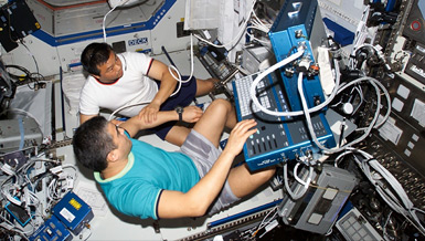
Dr. Yi-Xian Qin is developing an inflight device to monitor bone loss in both quality and quantity. The device, capable of generating noninvasive, high-resolution ultrasonic images of bones, can be used to determine subtle changes in density and strength of the skeleton in space and on Earth.
Overview
A Noninvasive Scanning Confocal Ultrasonic Diagnostic System for Bone Quality
Principal Investigator:
Yi-Xian Qin, Ph.D.
Organization:
Stony Brook University - State University of New York
Technical Summary
Currently funded by the NSBRI, we are able to develop a scanning, confocal, acoustic diagnostic (SCAD) system capable of generating acoustic images at the regions of interest (e.g., in the human calcaneus). This portable SCAD system is capable of generating noninvasive, high-resolution ultrasound (US) attenuation and velocity maps of bone, and thus determining the relationship between ultrasonic specific parameters, bone-mineral density (BMD), bone strength and bones physical properties (i.e., stiffness and modulus). This system is relevant not only for ground-based determination of bones physical properties, but can effectively be used in the space environment for determining even subtle changes in density and strength during extended flights.
In this study, we plan to develop a 2D ultrasound scanning system and validate the structure and density information detected by SCAD using CT and mechanical testing methods in ex vivo animal models as well as correlating to in vivo DEXA data, derived from humans. The system will thus contribute to monitor degree and risk of bone loss in space and Earth, as well serve as a major step towards clinical usage as an early diagnostic of osteoporosis.
There are proposed a series of four original specific aims (S.A.):
- Develop a scanning, confocal, acoustic diagnostic system for noninvasively mapping wave velocity and attenuation in bone;
- Determine an interrelationship between ultrasound-determined parameters, i.e., velocity and attenuation, and micro-architectural parameters in a quantitative manner;
- Develop a practical SCAD system for determining bone quality properties with quantified bone mass reduction, and;
- Map and monitor special directional and orthotropic strength of bone to predict BMD and structural modulus in vivo using the SCAD, and correlate these measurements to DEXA results.
During this award year (2002-2003), the research team focused on continuing technology development of SCAD system (S.A. 1 and 3) and validation between SCAD-determined acoustic parameters and bone quality data (S.A. 2). Human cadaver and in vivo subject testing were also initiated.
Technology Development
A system design including hardware and software of an experimental prototype was established and includes acoustic, electrical, control and mechanical components. As an important step towards a prototype for human testing, a 2D SCAD system has been built including converging ultrasonic transducers, micro-controller -controlled 2D scanning stages, ultrasonic wave generator, low-noise amplifier and real-time analog/digital (A/D) transformer. A specially designed controller was developed and synchronizes digital signals in acoustic wave, scan automation and A/D transform. This micro-controller -guided acoustic scanning technology greatly reduces the scan time, e.g., it requires only approximately three-to-four minutes for a 40x40 pixel array in the region of interest to form images with one-half to one -mm resolution. Ultrasonic attenuation and velocity images are obtained and calculated, e.g., gray scale or virtual color mapping.
SCAD as a noninvasive modality for animal bone-quality assessment: The ability of SCAD in noninvasively evaluating bone quality and quantity was tested in a large group of ex vivo bone samples. Trabecular samples were prepared as 1x1x1 cm cubes, which were harvested from sheep femoral distal condyle. These sheep were previously under a mechanical stimuli protocol for one-tp-two years and identified distinct bone-mineral density using dual-energy, X-ray absorptionmetry (DEXA). All bone samples were mechanically tested by direct-force deformation in orthogonal directions, i.e., longitudinal, medial-lateral and anterial-posterial, using a MTS universal test machine. The central plane of the samples was scanned with ultrasonic attenuation and velocity using SCAD system. The results of ultrasonic attenuation and velocity were correlated with mechanical moduli of the sample. While using a single transmitted ultrasound signal, there were weak correlations between measured BUA and micro-CT (CT) -determined osteo-parameters, e.g., BMD (R2=0.28), porosity (R2=0.28), trabecular thickness (R2=0.04) and trabecular space (R2=0.56), as well as average modulus (R2=0.40). These correlations were significantly improved using the SCAD system. Using the SCAD system, CT and mechanical testing, new constitutive relations were derived using linear regression correlations in the results, which predict BMD and bone stiffness as the functions of acoustic parameters using combined BUA and UV as well as a series of rational constants. Strong correlations are observed between SCAD-determined BUA and CT -determined parameters, i.e., BMD (R2=0.76), porosity (R2=0.61), structural mode index (R2=0.86), and average modulus (R2=0.71).
SCAD used for human calcaneus bone quality assessment: The feasibility of SCAD assessment for bone quality in the real body region, which includes soft tissue, cortical bone and different surface morphology, is evaluated in cadaver calcaneus. 19 human calcaneus, harvested from cadavers with ages from 66 to 97 have been imaged. BUA and ultrasound velocity determined from region of interest at the region of interest have been performed. Bone samples were further tested for structural and strength parameters using CT and mechanical testing in the extracted cylindrical samples (ten mm in diameter and 20 mm in medial-lateral length) from ROI. Strong correlations were found between BUA and bone volume fraction (BV/TV) (R2 = 0.76), and between UV and bones modulus (R2 = 0.53). The correlations are significantly improved (R2 > 0.64) using combined parameters of BUA and UV in linear regression, which ultrasound images determined parameters predicted structural properties, e.g., structure morphological index (SMI) (R2 = 0.86), and strength modulus (R2 = 0.64).
These results suggest that high-resolution acoustic mapping is capable of predicting calcaneus bone quantity and quality noninvasively. Structural property parameters of trabeculi, e.g., BMD and BV/TV, is better represented by BUA, while ultrasonic wave velocity has a strong agreement with bones strength property, e.g., modulus. Ultrasonic imaging has shown the great potential to be used as in vivo diagnostic modality for assessing skeletal disorder, i.e., osteoporosis.
Pilot study for human bone quality assessment at a large critical site, e.g., proximal femur, using SCAD and DEXA: To explore the potential of using SCAD to detect ultrasound bone image in the hip, the human-cadaver hip region is tested using an experimental SCAD system. This can evaluate the feasibility of ultrasonic assessment of bone quality in situ, which includes cortical bone and different surface morphology. The acoustic confocal region converged in the middle of the coronal plane of the hip with a focal zone approximately 0.5 mm in diameter in the focal region. Thus, a 2D scan covers the central bone of the proximal hip. The confocal scan area covers an approximate 100100 mm2 with a 0.5 mm increment. The signals transmitted through the bone are processed to calculate the slope of the frequency-dependent BUA (dB/MHz), the ultrasound attenuation (ATT, dB), and the ultrasound wave velocity (m/sec), and to generate BUA, ATT or UBV images. The data demonstrated that SCAD is capable of detecting bone tissues in the critical skeletal sites, e.g., the hip.
Impact
The results have demonstrated the feasibility and efficacy of SCAD for assessing bones quality in bone (CRL 4 and 5). With proof of the concept using SCAD in bone quality assessment, we have filed two new technology disclosures through Universitys Technology Transfer Office. Five journal papers are either published or under review, and approximately 12 conference papers were published, which are directly derived from this work. We have been able to demonstrate that the bone quality is predictable via noninvasive scan ultrasound imaging in the region of interest, and to demonstrate the strong correlation between SCAD-determined data and CT -identified BMD, structural index, and mechanical modulus. These data have provided a foundation for further development of the technology and the clinical application in this continuing research.
Research for the Coming Year
In the coming year, the research team will focus on 1). developing a practical SCAD system for determining bone quality properties with quantified bone mass reduction for clinical assessment, 2). assessing human calcaneus bone quality within a selected group, e.g., normal and osteoporosis subjects, to predict BMD and structural modulus in vivo using SCAD and DEXA, and 3). developing SCAD system for multiple sites testing.
A well-established SCAD system may provide a significant impact in diagnosis of osteoporosis and bone quality. Results may provide insight for addressing the risks of bone loss during prolonged space missions, age-related acceleration of osteoporosis and monitoring healing of fracture.


