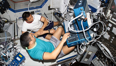
Analyzing blood composition is an important tool for monitoring health and making medical diagnoses. Presently, it is a slow process using bulky counting machines or trained technicians, and cannot be done in space. Tests from early missions showed that astronauts lost up to 20 percent of their red blood cell mass in just a few days in the weightless environment of space. On long missions, astronauts will need the ability to analyze blood samples to diagnose infection, allergies, anemia or deficiencies in the immune system. As such, Dr. Yu-Chong Tai is working to develop an automated, handheld blood-count machine that delivers immediate results and accurate readings. The project is to utilize the state-of-the-art microfluidics and lab-on-a-chip techniques so miniaturization of a blood count machine can be realized. He hopes to expand this first technology to analyze other body fluids such as urine, or to incorporate cell surface marker and DNA analysis.
Overview
Handheld Body-Fluid Analysis System for Astronaut Health Monitoring
Principal Investigator:
Yu-Chong Tai, Ph.D.
Organization:
California Institute of Technology
Technical Summary
During the previous funding year, we made progress in electrical impedance sensing. We solved a serious problem for micro-electrical impedance sensors, which is the large double layer impedance of electrode-electrolyte interface. The small surface area increases the double layer impedance and lowers the sensitivity. By either electroplating platinum black on the electrode surface or introducing an inductor parallel to the system to induce resonance, we achieve high signal-to-noise ratio when particles including blood cells pass the electrodes. At low AC frequency, the height of the signal was confirmed to be determined by the volume of the particles. The histograms of the signal height for whole blood and leukocyte-rich plasma matched very well to previous published volume distributions of erythrocytes and leukocytes. The inductor induced that a resonance-sensing approach can easily enable multi-frequency sensing by introducing multiple inductors with different inductance values to multiple pairs of electrodes along the same microfluidic channel. We successfully demonstrated fluorescent sensing and counting for WBC count and two-part differential with a portable prototype instrument. We established this method in previous funding years. In brief, cell nucleus fluorescent dye acridine orange stains DNA to be green and RNA to be red. Also, this dye is pH sensitive. As a result, non-lymphocytes have a higher red fluorescence level than lymphocytes due to their more active gene expression. The red fluorescence channel is used for the two-part WBC differential, while the green fluorescent channel is used for WBC count.
The unique feature of this technique is that whole blood can be used without dilution, which will greatly simplify system design, device operation and buffer storage. The concept was proved last year by constructing an optical detection system on an optical bench. Throughput of 1,000 WBCs/sec was achieved, which means a WBC count can be finished in one second. In this short period of time, blood cell sedimentation was not a problem. During the current year, we successfully made a portable prototype instrument based on this approach. The prototype instrument is made from off-the-shelf components with customer mechanical design. The system is composed of a blue LED for illumination, a fluorescent microscope filter block as main optical component, and two photon multiplier tubes for fluorescent signal detection. Data from the photomultiplier tube are amplified by a pre-amp and collected with a data acquisition board inside the instrument. The microfluidic chip is inserted into the sample chamber. A mini peristaltic pump is used to draw the sample through the device at up to 6 L/min. Typical time traces before and after the signal passing through a digital low pass filter were recorded. Two clusters corresponding to lymphocytes and granulocytes can be easily identified on the scatter plot. Comparison of lymphocyte percentages from ten clinical samples processed with the prototype and an automated hemacytometer resulted in good correlation.
During the past four years, we made significant advances on size-based microfluidic particle separation techniques with application on blood cell separation. Two main types of devices (pillar-shaped and channel-shaped) were demonstrated. RBC, WBC, and platelets can be separated into cell populations with over 96 percent purity. With the help of antibody conjugated beads, we also successfully achieved separation of subtypes of lymphocytes from the rest of the blood cells.
The findings above are key components to meet the specific aims of the proposal. Progress in electrical impedance sensing enables us to sense and count RBCs and WBCs with high signal-to-noise ratio, while separation of RBCs and WBCs makes the electrical impedance sensing more sensitive. We also showed under low AC frequency excitation, the signal height of the electrical impedance sensing was determined by the volume of the particle. So with proper calibration, the MCV could be measured directly by measuring individual cell volumes and averaging them. Hematocrit can be determined indirectly by adding volumes of individual cells together. Optical fluorescent sensing demonstrated two-part WBC differential from undiluted blood. Since fluorescent dye acridine orange and anticoagulant ethylene diamine tetraacetic acid can be coated on channel surface, sample preparation will be minimized, which will be very convenient for use in space. The system integration effort resulted in portable prototype equipment for WBC count and differential. This is the starting point for product development of the commercial hand-held hemocytometer.
This is the last year of the funding period for the current proposal. With the successful completion, we have recently started working on the continuation project. The new specific aims include:
- Five-part WBC differential;
- Analysis of WBC subtypes (e.g., CD4+ T helper and natural killer cells); and
- Serum/plasma protein biomarker analysis (e.g., for infection, radiation and bone loss monitoring).
Our proposed system has a much smaller size, lighter weight, lower voltage supply, less power and smaller sample volume. The functions provided by our proposed instrument are not available from any single commercially available instrument. More importantly, the proposed functions meet NASAs special needs for space missions.


