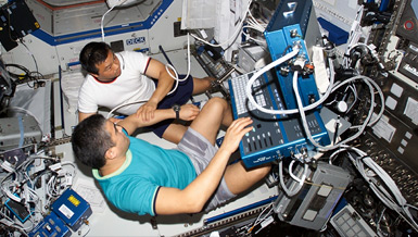
Impact of Long-Duration Spaceflight on Cardiac Structure and Function
Principal Investigator:
Zoran B. Popovic, M.D., Ph.D.
Organization:
The Cleveland Clinic Foundation
In order for humans to conduct long-duration missions in reduced gravity beyond low-Earth orbit, scientists must understand the impact of spaceflight on cardiovascular function. Dr. James D. Thomas is leading a project that will result in a comprehensive view of the heart in space.
To learn more about cardiac function in space, comprehensive analysis will be conducted on pre-, post- and in-flight cardiac studies over the next four years. The information will be added to mathematical models of the heart and made available to NASA researchers and physicians via the Digital Astronaut Project.
NASA Taskbook Entry
As astronauts venture farther into space, the impact of long-term microgravity on cardiovascular function may become a critical limitation to mission safety and success. In order to better understand the impact of long-term spaceflight on the structure and function of the heart, this project’s principal investigator, Dr. James Thomas, is already involved in echocardiographic analysis of the most detailed study of the heart in space ever undertaken. Unfortunately, the echocardiograph on the International Space Station is more than a decade old and does not provide contemporary information on cardiac function, such as strain (the best measure of regional and global contraction of the muscle) and torsion (the twisting motion of the heart that links the pumping and filling functions of the ventricle).
The first task in this project is to develop and validate methodology to extract strain and torsion from space station echocardiography and then combine it with the numerous pre- and post-flight studies that will be conducted over the next four years. From these data, the researchers will have a comprehensive view of the heart in space. This information will be integrated into evolving mathematical models of the heart developed by Thomas and his collaborators and will be made available to the NASA community via integration into the Digital Astronaut project. Finally, the researchers are involved extensively in the development of next-generation echocardiography machines and have the unique opportunity to develop and validate advanced applications for space use.
The project focuses on massively parallelized echocardiography machines capable of real-time 3-D imaging with automated volume measurements and comprehensive 3-D strain and torsion analysis. As these machines become smaller over time, they will provide the ideal diagnostic tool for future space missions, be they to low-Earth orbit, a Lagrangian point, the moon, or even Mars.
Exposure to microgravity induces short and long-term changes in the cardiovascular system, with cardiac atrophy, orthostatic hypotension and impaired thermoregulation being the most recognizable. The most obvious issue, noted in the majority of astronauts after long-term spaceflight, is orthostatic hypotension. While its importance is clear, the etiology remains uncertain, with proposed mechanisms including hypovolemia, impaired baroreflexes, and left ventricular atrophy leading to systolic and/or diastolic dysfunction. In order to better define these issues, NASA is currently conducting Flight Study E377, “Cardiac Atrophy and Diastolic Dysfunction During and After Long Duration Spaceflight: Functional Consequences for Orthostatic Intolerance, Exercise Capacity, and Risk of Cardiac Arrhythmias”. This program is also termed the Integrated Cardiovascular Study (ICV).
As part of this investigation, detailed imaging studies are conducted on astronauts before, during and after spaceflight, including an extensive series of in-flight resting and exercise echocardiograms. The PI monitors all in-flight echoes remotely in real-time, and he and his colleagues at the Cleveland Clinic serve as the echocardiographic core lab for this study. The researchers, thus, are in a unique position to guide on-flight acquisition as well as perform detailed examination of the ultrasound studies received. However, ICV was initially proposed in 1999 with echocardiographic techniques that are now more than 10 years old, focusing mainly on ventricular size, mass, and simple measures of systolic function, such as ejection fraction and stroke volume. Echocardiography has advanced considerably since then in the sophisticated ventricular mechanical data that can be extracted from ultrasound data. The researchers seek to validate extraction of these novel echocardiographic indices of ventricular mechanics (2-D strain and torsion, among others) from the in-flight data acquired on the 10-year-old HDI-5000 ultrasound system aboard the International Space Station, which was never designed to provide such data. Once validated, the researchers will be able to derive detailed regional ventricular mechanics from all of our in-flight studies, allowing direct comparison with the pre- and post-flight examinations to gain a much better understanding of the magnitude and time course of structural and functional changes in the cardiovascular system in microgravity.
These enhanced data from ICV will provide the ideal input for mathematical modeling of the cardiovascular system in space. The PI and colleagues have long experience with mathematical modeling ranging from lumped parameter models to 2-D structural models to full 3-D finite element models. The researchers will apply the structural and strain data from ICV to our evolving numerical models of the cardiovascular system. To model atrophy of the heart, The researchers will use the actual astronaut geometry from pre- and post-flight examinations, to build realistic 3-D finite element models. Chamber behavior will be extracted for use in our less computationally intense lumped parameter model. Such modeling will be made available to the NASA community to enhance the comprehensive Digital Astronaut model.
Finally, looking toward a future of long-duration missions to the moon and on to Mars, the researchers anticipate that even more sophisticated ultrasound data will be available through the expected commercial development of hand-held three-dimensional echocardiographs. The group stands in a unique position to capitalize on these developments and to validate their eventual use in the manned space program.
Several aspects of this project are already generating significant real-world benefits with many more anticipated in the future. Our work attempting to harmonize strain measurements across platforms has pointed out intervendor variability that significantly limits penetration of strain echocardiography into clinical practice. To address this, I have (in my role of President of the American Society of Echocardiography) convened a task force in collaboration with the European Association of Echocardiography and technical representatives from multiple vendors (GE, Siemens, Philips, Toshiba, Esaote, Ziosoft, Zonare, among others). We have proposed a multi-pronged validation protocol, consisting of synthetic datasets, animal experiments, and clinical validation at upcoming international congresses. In addition, we have engaged the DICOM (Digital Imaging and Communications in Medicine) committee with two proposals: 1) development of a new standardized format for storing raw ultrasound data (ideal for strain measurements) and 2) development of standardized nomenclature for advanced mechanics parameters, so analysis packages of the various vendors can communicate their results with each other and between data and picture archives.
Additionally, the modeling work being done in Cleveland and Auckland, while designed to allow simulation of the impact of physiological stressors in space flight on the cardiovascular system, will have widespread applicability in cardiology. For example, the user interface developed in Auckland allows any DICOM echocardiogram to be read into the program, segmentation and strain analysis to be performed, and then modeling of that heart using pre-existing fiber models of the ventricle. Once refined and validated, this should allow analysis of patients with regional and global dysfunction, as well as those with valvular heart disease. Work is also underway to allow 3D echoes to be read directly into the interface, including the full strain tensor as reported throughout the 3D space (Toshiba machine).
Finally, we have leveraged our work in 3D echocardiographic strain to participate in an international consortium to establish normal values for global and regional 3-D strain. Three-dimensional echocardiography is undergoing rapid development and this work will help to set normative standards against, which clinical acquisitions can be compared.


