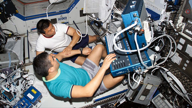
Skeletal complications, i.e., osteoporosis, induced by microgravity during extended space mission represent a key astronaut health problem. Lack of on-board diagnosis has increased significant risk in astronauts’ bone loss during long term space flight. Early diagnosis of such disorders can lead to prompt and optimized treatment that will dramatically reduce the risk of fracture and longitudinal monitoring microgravity and countermeasure effects. Advents in quantitative ultrasound (QUS) techniques provide a method for characterizing the material properties of bone in a manner for predicting both BMD and mechanical strength. We have developed a scanning confocal acoustic navigation (SCAN) system capable of generating noninvasive ultrasound images at the site of interest. Both animal and human tests indicated strong correlations between SCAN determined data and microCT determined BMD, and bone strength, as well as monitoring fracture healing with guided ultrasound. The objectives of this study are to develop a portable broadband SCAN for critical skeletal quality assessment, to longitudinally monitoring bone alteration in disuse osteopenia, and to integrate ultrasound with DXA, QCT and finite element analysis (FEA) for human subject. In vivo human tests will be evaluated at Stony Brook Osteoporosis Center. Human cadaver will be used for testing feasibility of identifying bone loss, microstructural and mechanical strength properties. Development of a noninvasive diagnostic and treatment technology using noninvasive ultrasound with new crystal transducer technology will have a great potential to perform longitudinal measurement of bone alteration and prevent the risk of fracture.
Overview
Portable Quantitative Ultrasound with DXA/QCT and FEA Integration for Human Longitudinal Critical Bone Quality Assessment
Principal Investigator:
Yi-Xian Qin, Ph.D.
Organization:
SUNY - The State University of New York
Technical Summary
Non-invasive assessment of trabecular bone strength and density is extremely important in predicting the risk of fracture in space and ground operation. Quantitative ultrasound (QUS) has emerged with the potential to directly detect trabecular bone strength. To overcome the current hurdles such as soft tissue and cortical shell interference, improving the "quality" of QUS and applying the technology for future clinical applications, the development of image based SCAN system will concentrate on several main areas: (1) increasing the resolution, sensitivity, and accuracy in diagnosing osteoporosis through confocal acoustics to improve signal/noise ratio, and through extracting surface topology to accurately calculate ultrasound speed of sound; (2) minimizing the scanning time while maintaining reasonable resolution via micro-processor controlled and phased array electronic confocal scanning, e.g., in deep bone tissue scan; (3) developing broadband ultrasound system to measure deep bone tissue density and structural parameters in the critical regions; (4) validation of structural and strength properties using micro-CT, nanoindentaiton and mechanical testing, and DXA; (5) feasibility assessment in human subject testing; and (6) guided treatment for early fracture using ultrasound. The proposed work will develop a portable rapid SCAN system combined with imaging capability, and test its efficacy in human, which will ultimately provide a portable, noninvasive device for bone loss assessment in space.
Earth Applications
Skeletal decay due to a microgravity environment has greatly impacted the nation's civil space missions and ground operations. Such musculoskeletal complications are also major health problems on Earth, i.e., osteoporosis, and delayed healing of fractures. Development of a low mass, compact, noninvasive diagnostic and treatment technology, i.e., using ultrasound, will have a great potential to prevent and treat bone fracture. Our principal goal is to develop a portable quantitative ultrasound system with therapeutic capability, not only for determination of bone's physical properties, but also for predicting subtle changes of bone during extended flights and diseased condition, which will impact both diagnosis and noninvasive treatment for musculoskeletal disorders.


