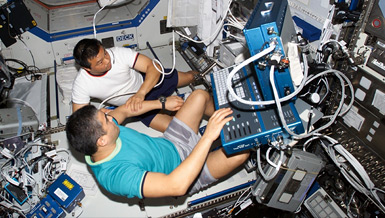
In NASA space missions about 15% of astronauts are women, and women made up half of the 2013 NASA Astronaut Class. However, risks of uterine exposure to galactic cosmic rays and solar particle events during space missions remain completely unknown. Normal uterine structure and function is required for a healthy pregnancy and optimal development and subsequent health of the offspring. Uterine function in adults is largely regulated by ovarian steroid hormones secreted from the growing pool of follicles. Our preliminary data generated under a NASA pilot grant showed that Iron charged particle radiation induces apoptosis and destroys ovarian follicles in mice. Radiotherapy for the treatment of cancer in the pelvic region has been shown to damage uterine cells, reduce blood circulation to the uterus, and increase the risk for miscarriage, low-birth weight, and premature birth. In addition, pelvic irradiation for cancer treatment is associated with higher risk for uterine cancer. Female atomic bomb survivors have increased risk of uterine leiomyoma. Therefore, this project focuses on the risk of space radiation on uterine health.
Overview
Effects of charged particles on the uterus (First Award Fellowship)
Principal Investigator:
Birendra Mishra, Ph.D.
Organization:
University of California, Irvine
Technical Summary
Proper functioning of uterine cells is essential for embryo implantation and placental development, which includes the proliferation of luminal and glandular epithelial cells, decidualization of stromal cells and angiogenesis. Based on the histological observations of early cell death, the uterus is highly sensitive to ionizing radiation and is considered as group-II of the classification of tissue radiosensitivity of Casarett. In premenopausal women, radiation therapy has been shown to induce a decrease in uterine size, uterine thickness, and myometrial relaxation after 3-6 months of therapy. In childhood cancer survivors, radiotherapy also causes premature ovarian failure, and decreases endometrial thickness in adult life. Recently, flattening of endometrial surface, depletion of deep glands and reduced mitotic figures were reported in rats irradiated with 5 Gy of Cesium-137. However, effects of high charge and energy (HZE) charged particles on the uterus have not been examined. Based on epidemiological data on earth and our preliminary findings generated under the NASA pilot grant, we therefore hypothesize that exposure to HZE particles typical of space radiation alters uterine cellular functions, pregnancy outcomes and also contributes to pathogenesis of uterine cancer and leiomyoma. These hypotheses will be tested in the following specific aims: 1) Acute exposure to charged particles induces uterine oxidative stress, 2) Acute exposure to charged particles alters pregnancy outcome, 3) Exposure to charged particles causes uterine cancer and leiomyoma.
Aim 1: Acute exposure to charged particles induces uterine oxidative stress.
This aim will examine the effects of charged particles on the various uterine cell types; luminal and glandular epithelium, stromal cells and endothelial cells which are important for the outcome of pregnancy. Uterine samples were collected from three month old female mice (C57BL/6J) exposed to low dose (0, 5, 30 and 50 cGy; n=8/treatment) iron (LET = 179 keV/µm) at an energy of 600 MeV/u. Two groups were irradiated at the highest dose for each of the two charged particles, one fed AIN-93M chow and the other fed the same chow supplemented with 150 mg/kg chow of the antioxidant alpha lipoic acid began one week before irradiation and continued until euthanasia. Uteri were fixed in 4% Paraformaldehyde and embedded into OCT. To study the uterine morphology, sections will be stained with Hematoxylin and Eosin. To understand the mechanisms by which charged particle irradiation induce damage to uterine tissues, markers of DNA double strand breaks (ƴH2AX), apoptosis (activated Caspase-3), oxidative damage for protein (NTY) and Lipid (4-HNE), cell proliferation (PCNA) and Ki-67 will be analyzed.
Aim 2: Acute exposure to charged particles alters pregnancy outcome.
This aim will examine the effects of charged particles on pregnancy outcome using adult mice. Embryo implantation to uterus is a highly complex and dynamic process, which involves an intricate discourse between the embryo and uterus. Uterine stromal cells that surround the blastocyst undergo decidualization following attachment, eventually embedding the embryo in the stromal bed. Several epidemiological studies have demonstrated an increased risk of adverse pregnancy outcomes in women previously exposed to childhood abdominal radiation and/or total body irradiation. There is no information in the literature about the fertility and pregnancy outcome in women who have been exposed to pelvic radiation in adulthood. Atomic bomb exposed women who married unexposed males had 1.4 births each between 1948 and 1954 in Japan. In the NASA corps, the average age of women astronauts at selection is 32.8 (26-47) years; indicating that they still have reproductive years after the space mission.
Three month old female mice (C57BL/6J) will be exposed to low dose (0 and 50 cGy; n=8/treatment) iron (LET = 179 keV/µm) at an energy of 600 MeV/u. After irradiation, female mice will be shipped to UC Irvine and acclimatized for two weeks. These female mice will be mated with fertile males of the same strain to induce pregnancy. Mating of mice will be monitored daily in the morning for the presence of vaginal plug (day 1 of gestation = the day of vaginal plug). The implantation sites on day 6 (DPC-6) of pregnancy will be identified by intravenous injection of 1 ml of 1% Chicago blue (Sigma) in 0.85% sodium chloride. Mice often cannibalize their pups immediately after birth, therefore, the number of fetuses will be determined at day 18 of pregnancy (DPC-18, close to delivery). Uterine tissues will be collected to analyze the markers of uterine remodeling (EMMPRIN) and angiogenesis (VEGF). Ovaries from these mice will also be fixed in Bouins solution, and embedded in paraffin. Ovarian sections will be stained with Hematoxylin and Eosin to count the number of corpora lutea.
Aim 3: Exposure to charged particles causes uterine cancer and leiomyoma.
Uterine cancer is the fourth most common cancer and the seventh most common cause of cancer death for women in the United States. Based on uterine cancer patient history, 55% were nulliparous and 39% reported irregular menstrual cycles suggesting that alterations in reproductive cycles is one of the predisposing factors for uterine cancer. During long duration spaceflight, female astronauts will spend a longer period of time in space and they will not have regular menstrual cycles, which may increase their risk of uterine cancer in and of itself and by interacting with space radiation exposure. Uterine tumor has also been reported in women several months after irradiation for pelvic malignancy. About 20% of the females exposed to atomic bomb radiation were found to have uterine nodules; known as myoma, fibroid, or leiomyoma. Although initiators of leiomyoma are not fully identified, estrogen and progesterone are known to promote leiomyoma directly or through regulating cytokines, chemokines and growth factors. Several groups found that expression of estrogen receptors is higher in leiomyoma compared with control myometrium. Estrogen binds with its receptor and stimulates the proliferation of leiomyoma cells by activating growth factors, and suppressing apoptosis-specific signaling molecules (p53). Our preliminary data showed that the majority of ovarian follicles are depleted after HZE irradiation. It has also been shown that the remaining theca cells of depleted ovarian follicles produces androstenedione; a precursor of estradiol. Aromatase, the enzyme that catalyzes the final step in the synthesis of estradiol from androstenedione is over expressed in leiomyoma cells. So, we speculate that myometrial aromatase will convert androstenedione secreted from theca cells of the ovary, which will promote leiomyoma. Therefore, under this aim we will test the hypothesis that HZE irradiation induces uterine cancer and leiomyoma. Three month old female mice of two strains, one thought to be sensitive to radiation induced tumors (B6C3F1) and one thought to be less sensitive to radiation-induced tumors (C57BL/6J) were exposed to low dose (0 and 50 cGy; n=15/treatment) iron (LET = 179 keV/µm) at an energy of 600 MeV/u. Uteri were collected at 15 months after irradiation. These tissues were fixed with 4% PFA or 10% neutral formalin. For histomorphological study, uterine sections will be stained with Hematoxylin and Eosin. For detection of endometrial tumor; CA-125 (cancer antigen 125) and Ki67 immunostaining will be done. EMMPRIN is highly expressed in tumor tissues and stimulates the expressions of several metalloproteinase which in turn degrade the extracellular matrix for tumor cell metastasis. To study the metastasis of tumor in the uterus, expression of EMMPRIN will be analyzed. Over expression of estrogen receptor, and mutation in MED12 are associated with leiomyoma. In order to confirm leiomyoma, expression intensity of estrogen receptor and MED12 will be examined by immunohistochemistry.
Earth Applications
The findings of this study will not only provide the risk of space radiation on reproductive health of female astronauts but also fill the gaps of knowledge on the earth. There is no information in the literature about the fertility and pregnancy outcome in women who have been exposed to pelvic radiation in adulthood. Uterine sensitivity to ionizing radiation has primarily been studied using low linear energy transfer (LET) radiation such as x-rays, and gamma rays which are used for radiotherapy. High LET ions such as protons and carbon ions are increasingly being used for cancer treatment, and these charged particles are also constituents of galactic cosmic rays. Therefore, the findings of this study also provide the basic information of uterine risk of charged particle radiation. If the supplementation of alpha lipoic acid mitigate the radiation effects on uterus, it could be used as therapeutic prevention in the patient before undergoing radiotherapy.


